No, teeth cannot grow out of your toes. There is no credible medical evidence that true tooth tissue, meaning enamel, dentin, or any part of an actual tooth, can develop and erupt from toe tissue in humans. If you're seeing something hard, sharp, or tooth-like on your toe right now, it is almost certainly something else entirely: a bone spur, a wart, a callus, a nail abnormality, or in rarer cases a tumor or infection. The good news is that all of those things have real diagnoses and real treatments, and a doctor or podiatrist can figure out exactly what's going on.
Can Teeth Grow Out of Your Toes? What to Know and Do

Marcus Holloway
18 May 2026
Why teeth literally cannot form in your toes
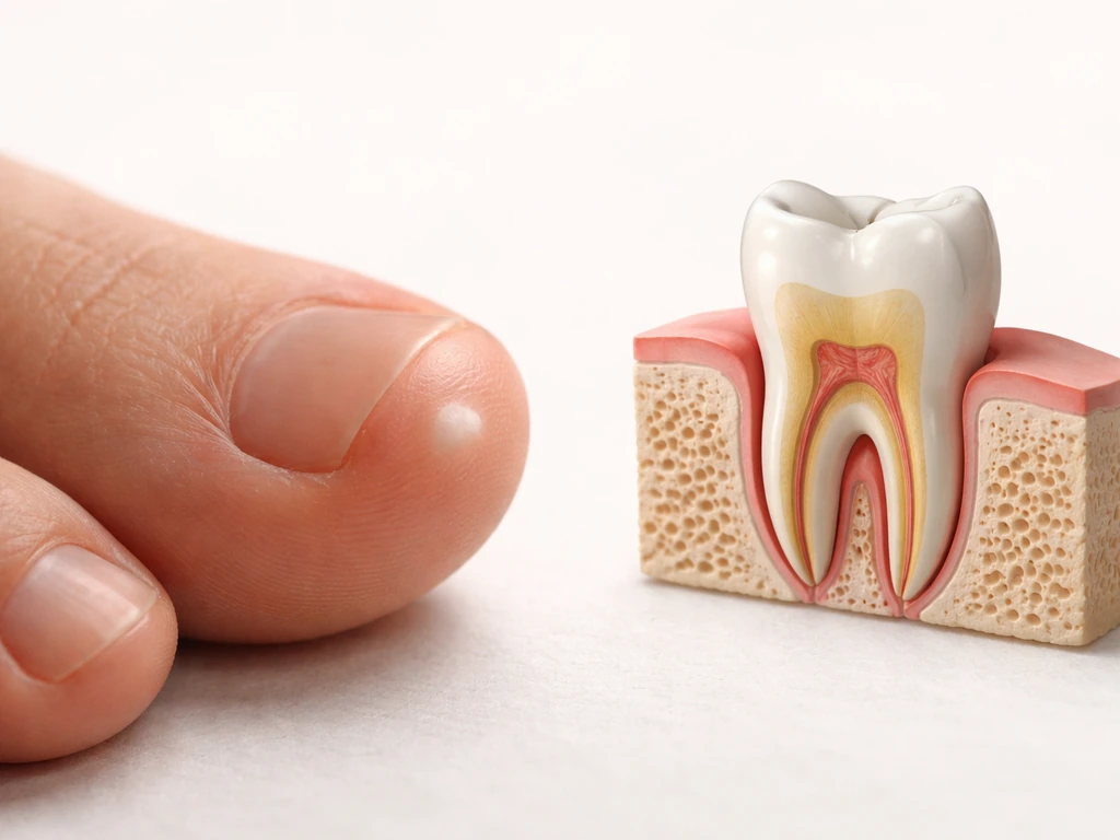
Tooth development is one of the most complex and tightly controlled processes in the human body. It doesn't just happen anywhere there's tissue. To form a tooth, you need a very specific combination of oral epithelium (the lining of the mouth) and neural crest-derived mesenchyme (specialized cells that migrate from the developing brain region during embryonic development). These two cell types interact through a coordinated signaling program involving FGF and BMP family molecules, eventually producing the enamel-forming cells called ameloblasts and the dentin-forming cells called odontoblasts.
That entire cascade starts in the jaw during embryonic development, organized around a structure called the dental lamina. Without a dental lamina, without the right epithelial-mesenchymal interaction, and without the oral epithelium that's been programmed to respond to those signals, a tooth simply cannot form. The skin, connective tissue, and bone of your toes have none of those components. They are not even close to the right environment. This is why tooth development in adult mammals essentially does not happen outside the jaw, and why researchers trying to bioengineer replacement teeth have to work so hard to reprogrammatically induce tooth-forming potential in non-dental cells.
There's also a developmental timing element. Tooth formation happens during a specific embryonic window, and the signaling centers that guide it, including structures called enamel knots that shape cusp patterns, are transient. They appear, do their job, and shut down. There is no mechanism sitting dormant in your toes waiting to restart that process.
What people actually see on their toes (and mistake for something tooth-like)
The real question here is: what are you actually looking at? Several conditions can produce hard, pale, protruding, or sharp structures on or around the toes that might prompt the thought. Here are the most likely culprits.
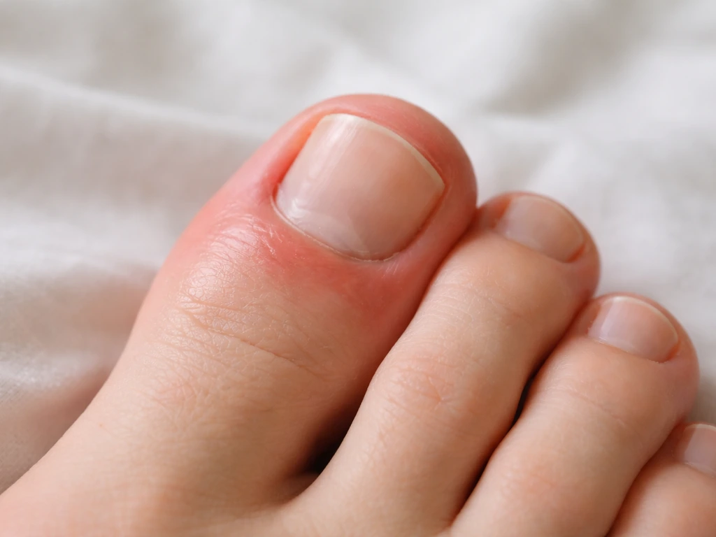
Subungual exostosis (bone spur under the nail)
This is probably the most common reason someone would describe something that feels genuinely tooth-like on a toe. Subungual exostosis is a benign outgrowth of bone or calcified cartilage from the tip of the toe, usually the big toe. It grows up under the nail bed, lifting or separating the nail, and presents as a firm, flesh-colored or pinkish nodule that can reach 8 to 10 mm in diameter. It is hard as bone because it is bone. It can be painful, especially when pressure is applied. An X-ray confirms the diagnosis immediately, because the bony origin shows up clearly on imaging.
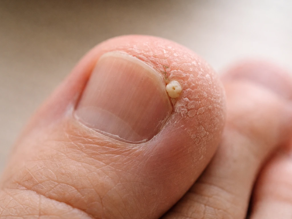
Plantar warts and verrucas
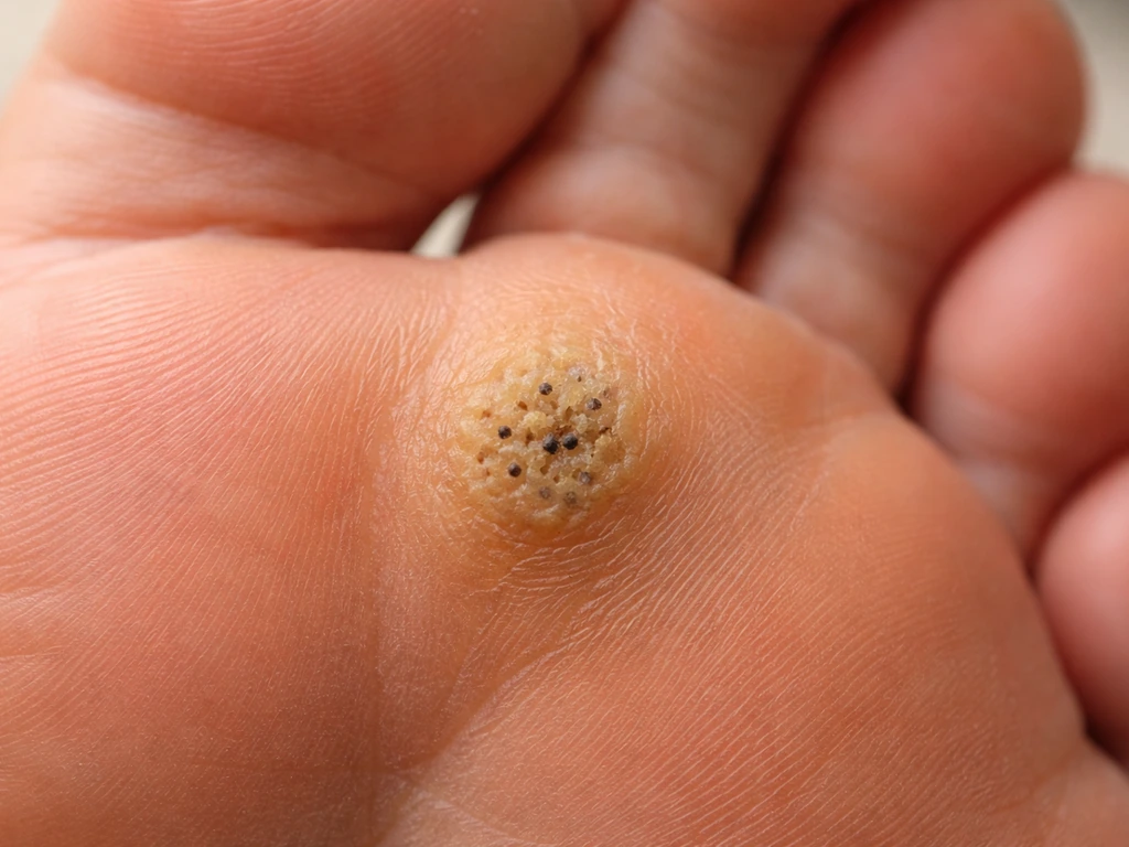
Plantar warts are caused by human papillomavirus (HPV) infecting the skin of the foot. They can appear on the toes and often have a roughened, cauliflower-like surface with distinctive black dots inside, which are actually thrombosed (clotted) capillaries, not anything structural. Warts can vary in color from white and pink to brown, gray, or black. They tend to hurt when you press directly on them or when walking puts weight on them. A dermatologist can distinguish warts from other lesions using dermoscopy, which reveals the characteristic papillary capillary pattern of viral warts.
Calluses and hardened skin
Repeated friction or pressure causes the skin to produce thick, hardened layers of keratin as a protective response. On toes, calluses can build up to become genuinely hard, pale, and dense. They don't have blood supply running through them, which is part of why they can look almost structural. Calluses are not painful on their own but may cause discomfort when they press on underlying tissue. They're completely benign and usually resolve with moisture, padding, and pressure relief.
Nail abnormalities and onychomycosis
Fungal nail infections (onychomycosis) cause nails to thicken, become discolored (yellow, white, brown), and take on a crumbly, distorted texture. In advanced cases, the nail can separate from the bed and develop irregular edges or protrusions that feel sharp. This is purely a nail and skin issue, not anything involving bone or tooth tissue. It's also extremely common, affecting roughly 10% of the general population.
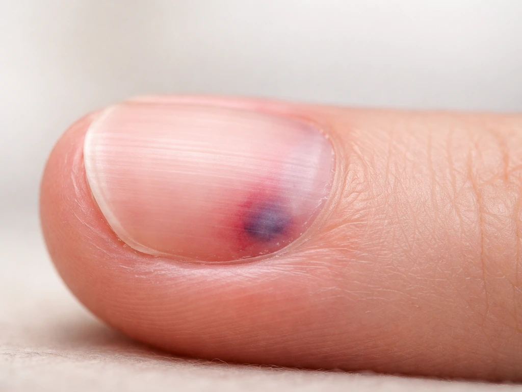
Glomus tumor
Less common but worth knowing about: a glomus tumor is a small, benign vascular tumor that can grow under or around the nail on a finger or toe. It presents as a tiny reddish-blue papule or nodule and is known for causing severe, disproportionate pain, especially with cold exposure or light touch. The classic triad is pinpoint tenderness, cold sensitivity, and a small visible lesion. If your toe pain is way out of proportion to what you can see, a glomus tumor is worth ruling out.
Infection and cellulitis
Bacterial skin infections can cause swelling, redness, and tissue changes that produce unusual textures or protrusions around toes, especially near the nail folds. Cellulitis involves redness, warmth, swelling, and pain. If redness is spreading quickly or you have fever and chills alongside a toe problem, that's a medical urgency, not something to watch at home.
How to assess what you're seeing right now
Before you call anyone or search anything else, take a few minutes to observe the lesion carefully. Here's what to look for and what each clue means:
| What you notice | Most likely cause | Urgency |
|---|---|---|
| Hard, firm nodule lifting the toenail, no color change | Subungual exostosis (bone spur) | See podiatrist or GP soon, not emergency |
| Rough, cauliflower-like surface with black dots | Plantar wart | See dermatologist or GP, not urgent |
| Thick, yellowed, crumbly nail with distorted edges | Fungal nail infection | See GP or dermatologist, not urgent |
| Extreme pain from light touch or cold, reddish-blue spot under nail | Glomus tumor | See GP or dermatologist soon |
| Dark streak or band under nail, growing toward cuticle | Possible subungual melanoma | See doctor promptly, do not delay |
| Redness spreading up foot, warmth, fever, chills | Cellulitis (bacterial infection) | Seek urgent medical care today |
| Pale, thick, hardened skin patch, not painful | Callus | No urgency, routine care |
One condition deserves a special mention: subungual melanoma. It can start as a small dark streak or band under the nail, resembling a bruise, and gradually expand to cover more of the nail and extend toward the cuticle. It affects the digits, including the great toe, and is sometimes mistaken for a fungal infection or injury. If you're seeing a dark discoloration under a toenail that isn't explained by a recent injury and isn't improving, get it checked by a doctor promptly. This one is not something to wait on.
What to do next: home care and when to get professional help
For most of the benign causes above, there are sensible home steps you can take while arranging to see someone if needed.
- Photograph the lesion now and again in a few days to track whether it's growing, changing color, or changing shape. This is genuinely useful information for a clinician.
- Avoid cutting, picking, or scraping the area, especially if it's under the nail or near the nail fold. You can introduce infection or make diagnosis harder.
- If it's a callus or rough skin, soak the foot in warm water for 10 to 15 minutes, then gently use a pumice stone. Moisturize afterward. If it doesn't improve within two to three weeks, see someone.
- If it's painful, wear footwear that reduces pressure on the area. Cushioned insoles or toe padding can help.
- If there's any redness, warmth, pus, or spreading discoloration, stop home management and call your GP or visit urgent care the same day.
- If you see a dark streak under the nail with no known cause, book an appointment with your GP or a dermatologist this week, not eventually.
In terms of which specialist to see: a podiatrist is your best first call for anything structural involving toes, nails, or foot bones (bone spurs, nail problems, calluses). A dermatologist is the right choice for skin lesions, warts, or anything involving pigmentation or nail discoloration. Your GP can coordinate either referral and can also rule out infection or systemic causes. If the lesion is hard and bony, ask for an X-ray, which is the standard first imaging step for confirming subungual exostosis.
The truth about dental regrowth (since that's why you're here)
Since this site focuses on whether teeth and dental structures can actually grow back, it's worth being direct about what the biology allows, because there's a lot of confusion out there.
Enamel is the hardest substance in the human body, and it cannot regenerate. Once it's gone, it's gone. The cells that produce enamel, called ameloblasts, do their job during tooth development and then die off or withdraw before the tooth even erupts. After that, there are no living enamel-producing cells left in your tooth. What is possible is remineralization, which means using minerals (typically calcium and phosphate, sometimes fluoride or casein phosphopeptide-amorphous calcium phosphate, known as CPP-ACP) to partially repair early surface damage and strengthen weakened enamel. This is not growing enamel back; it's shoring up what remains. Studies evaluating remineralization agents at 7, 14, 21, and 28-day time points confirm measurable mineral deposition, but it doesn't replace bulk enamel lost to decay or wear.
Dentin has slightly more potential because the dental pulp contains stem cells that can produce some secondary or tertiary dentin in response to injury. But that capacity is limited and depends on a healthy, infection-free pulp. Cementum (the tissue covering tooth roots) also has limited regenerative capacity in the context of periodontal disease. Importantly, natural tooth eruption only happens in the jaw during defined developmental windows: primary (baby) teeth and then permanent teeth. After that, the system is done. There's no third set waiting in the wings, regardless of what some internet claims suggest.
Regeneration vs. restoration: what's actually available today
If you're interested in the regeneration side of things because you've lost tooth structure, here's a realistic picture of where things stand in 2026.
- Remineralization (toothpastes with fluoride, CPP-ACP products, professional fluoride treatments): Works for very early enamel lesions and surface weakening. Takes weeks to months of consistent use. Does not replace lost enamel.
- Periodontal regenerative surgery (guided tissue regeneration, enamel matrix derivatives): Can restore some lost bone and soft tissue support around teeth affected by gum disease. Systematic reviews confirm efficacy for early wound healing outcomes. Timelines are typically measured in months for initial healing, with continued remodeling over a year or more.
- Dental restorations (fillings, crowns, veneers, implants): These are the practical, proven options for restoring lost tooth structure or replacing missing teeth today. Implants integrate with jaw bone over three to six months.
- Tooth bioengineering research: Scientists are working on techniques to grow whole teeth from stem cells, but this requires reprogramming non-dental cells to acquire tooth-forming capacity. It is not clinically available yet, and realistic timelines for human applications remain unclear.
The pattern with dental regeneration is consistent: natural regrowth is extremely limited, especially for enamel, and current solutions are restorative rather than truly regenerative. Researchers are making progress, but the biological hurdles are significant precisely because tooth formation is such a tightly controlled developmental process.
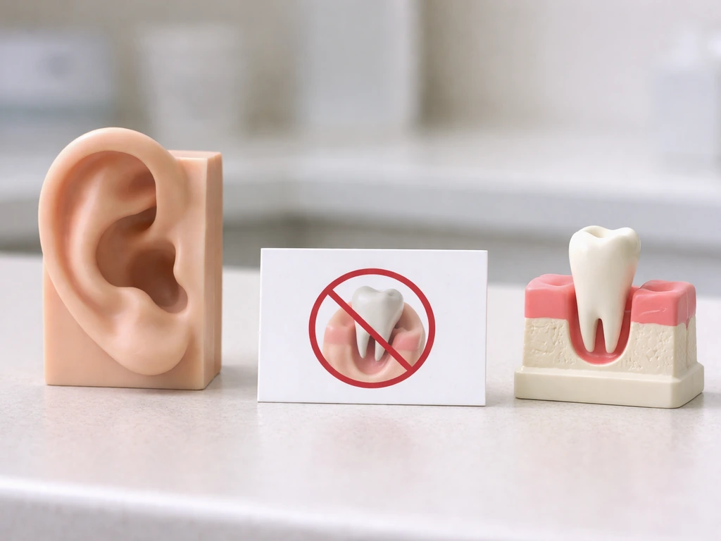
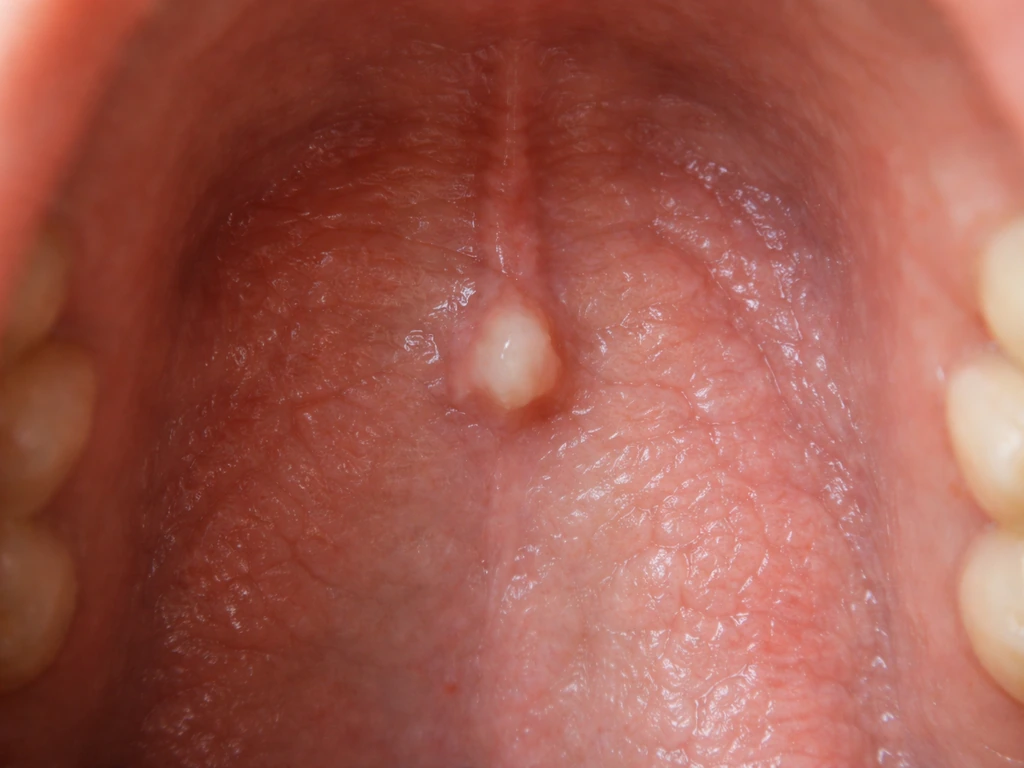
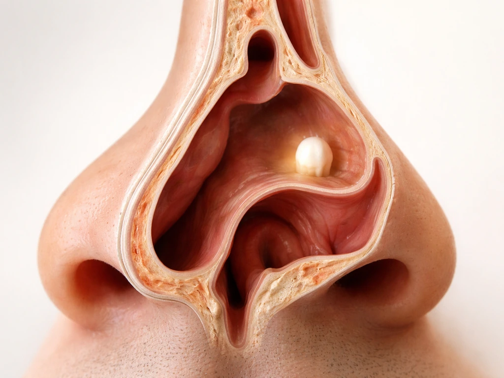
It's worth noting that the question of teeth appearing in unexpected locations isn't unique to toes. Similar curiosity comes up around teeth found in the nose, on the roof of the mouth, in the ear canal, and in the throat. So, can a tooth grow in your nose? A tooth cannot grow in your throat either, because the same jaw-based developmental program is required for true tooth tissue to form can a tooth grow in your nose. It is also not possible for true tooth tissue to grow in your ear for the same developmental reasons. In most cases, anything that looks tooth-like is misidentified, because true tooth tissue forms only with the jaw-specific developmental program teeth found in the nose. In all of those cases, the biology is the same: true tooth tissue requires the specific developmental program of the jaw, and anything found elsewhere is either misidentified, a rare developmental anomaly (like a teratoma containing tooth-like structures formed during fetal development), or simply a completely different type of lesion. None of those situations involve a tooth spontaneously growing where it shouldn't.
Bottom line: if something on your toe looks or feels strange today, get it properly diagnosed. It won't be a tooth, but it might be something that genuinely needs attention, and knowing what it actually is will point you straight to the right treatment.
FAQ
What does a toe lesion have to look like before I should worry it could be something serious, not a callus or wart?
Get it checked promptly if it is rapidly growing, bleeding without a clear injury, has a dark color that is changing, or causes severe pain that feels out of proportion to what you can see. Also seek care sooner if you have fever, spreading redness, or trouble walking due to swelling.
Can a fungus infection or ingrown toenail cause something that feels hard or tooth-like?
Yes. Advanced fungal nail disease can distort the nail so it becomes thick and sharp at the edges, which can feel like a hard protrusion. Ingrown nails can also create firm, painful tissue around the nail fold. In both cases, the “hard spot” is coming from nail and skin tissue, not enamel or dentin.
Are black dots on a toe always warts?
Not always. Black dots can be thrombosed capillaries and are typical of plantar warts, but other issues, including some pigmented lesions or infections, can also darken. The quickest way to tell is a clinician exam (sometimes dermoscopy) rather than trying to match images online.
If it hurts when I press on it, does that point more toward a wart or a bone spur?
Pain with direct pinpoint pressure can fit either, but subungual exostosis tends to feel like a fixed, bony nodule under or near the nail and is confirmed with an X-ray. Plantar warts often hurt when you press and when walking applies weight, and they typically have a rough surface with internal black dots.
Should I try trimming it, picking at it, or using wart removers on my toe?
Avoid cutting, digging out, or aggressive trimming. If the cause is an exostosis, cutting can worsen nail lifting or increase injury. For suspected warts, OTC treatments can be slow and not ideal if the lesion is actually something else, like a nail condition or a pigmented lesion, so it is safer to get diagnosed first when the appearance is unclear.
How do I know whether I need an X-ray?
Ask for imaging if the growth feels hard like bone, is under the nail, or you notice nail lifting with a firm protrusion. An X-ray is the standard first step to confirm subungual exostosis. If redness and warmth suggest infection, prioritize urgent clinical evaluation rather than imaging at home.
Which specialist should I see if the problem is both skin and nail, like discoloration plus a hard bump?
Start with a podiatrist for structural toe or nail issues, especially if there is a firm, bony-feeling bump or nail changes. See a dermatologist if the main problem is a skin lesion, wart-like growth, or significant pigmentation changes. A primary care clinician can triage and coordinate referrals if you are unsure.
What is the key warning sign for subungual melanoma compared with a bruise or fungal nail issue?
A main red flag is a dark discoloration under the nail that does not have a clear injury history and does not steadily improve over time. Worrisome cases may expand toward the cuticle or change in width. Prompt evaluation matters because early diagnosis changes outcomes.
Can tooth-like material on a toe be a tumor or infection?
Yes, though less commonly. Glomus tumors can cause intense, cold-sensitive pain with a small visible lesion, and infections can cause swelling, warmth, redness, and tissue changes. If you have spreading redness, fever, or chills, treat it as urgent rather than trying home remedies.
If enamel cannot regenerate, can dental treatments “grow” enamel or teeth elsewhere on the body?
No. Enamel cannot regenerate because the enamel-forming cells are not available after tooth development. Dental therapies like remineralization can strengthen early surface damage, but they do not replace lost bulk enamel, and true tooth formation does not occur outside the jaw developmental program.
Citations
There is no credible medical evidence that true, naturally forming tooth tissue (enamel/dentin) can develop and erupt from human toes (non-jaw sites); when people report “tooth-like” material on a toe, clinicians typically find a non-tooth cause (e.g., skin/nail lesions, bone outgrowth, or other growths) rather than odontogenesis.
https://www.ncbi.nlm.nih.gov/books/NBK557543/
In humans, tooth development requires a specialized tooth-forming organogenesis program involving the dental lamina and epithelial–mesenchymal interactions; tooth eruption is described as penetration into the oral soft tissue after tooth development in the jaw.
https://www.ncbi.nlm.nih.gov/books/NBK557543/
Known tooth development involves ectoderm-derived oral epithelium forming the enamel-producing lineage and neural crest ectomesenchyme contributing other tooth structures; tooth formation is described as a complex process requiring these interactions.
https://www.ncbi.nlm.nih.gov/books/NBK560515/
Tooth organogenesis depends on dental lamina formation (an epithelial thickening at the future tooth site) and differentiation at the epithelial–mesenchymal junction: enamel is produced by ameloblasts and dentin by odontoblasts.
https://www.ncbi.nlm.nih.gov/books/NBK27071/
During crown formation, enamel knots serve as a signaling center that mediates differentiation of odontoblasts and ameloblasts and patterns tooth cusps; this is part of the normal jaw/tooth developmental program.
https://pubmed.ncbi.nlm.nih.gov/30902260/
Experimentally and developmentally, tooth-forming competence involves specific signaling pathways (including FGF and BMP family signaling from oral epithelium to underlying mesenchyme) used to pattern tooth fields in the developing mouth.
https://www.nature.com/articles/nrg1380
Tooth-like structures are often misidentified clinically as dermatologic or nail disorders; for example, plantar warts can show “black dots” (thrombosed capillaries) and can be painful on weight-bearing pressure.
https://my.clevelandclinic.org/health/diseases/24899-plantar-warts
Dermatology guidance describes wart appearance variability (colors including brown/gray/black/white/pink) and notes black dots in warts can represent blood vessels.
https://www.aad.org/public/parents-kids/healthy-habits/parents/kids/warts-look-like
Warts can be distinguished with dermoscopy: dermoscopy can visualize papillary capillaries of viral warts and help distinguish verrucous lesions from other skin conditions.
https://dermnetnz.org/topics/viral-wart
Subungual exostosis is an outgrowth of bone from the tip of the toe; it presents as a firm nodule below the nail bed and nail plate separation (onycholysis) with often pain as it grows.
https://dermnetnz.org/topics/subungual-exostosis
Subungual exostosis size and diagnostic approach: one source describes a solitary pink/flesh-coloured firm swelling below the nail that may reach ~8–10 mm diameter and that X-ray examination confirms bony origin.
https://www.dermis.net/dermisroot/en/20787/diagnose.htm
Subungual exostosis diagnosis is confirmed with imaging such as X-rays; imaging is emphasized because the lesion is bony rather than a skin-only growth.
https://dermnetnz.org/topics/subungual-exostosis
Glomus tumor (a painful digit tumor) can occur under or around the nail and may present as a solitary painful reddish-blue papule/nodule; key features include severe localized pain and nail-bed color change in some cases.
https://dermnetnz.org/topics/glomus-tumour
A review article on glomus tumors notes the “classic triad” involves pain/pinpoint tenderness and cold sensitivity; diagnosis is clinical with history/physical maneuvers.
https://pmc.ncbi.nlm.nih.gov/articles/PMC4567371/
Cellulitis warning signs: CDC notes cellulitis is bacterial infection causing redness, swelling, and pain; urgent medical attention is advised if redness spreads quickly or fever/chills occur.
https://www.cdc.gov/group-a-strep/about/cellulitis.html
NHS guidance for cellulitis: advises urgent GP appointment or NHS 111 if cellulitis is suspected and highlights that untreated infection can spread to other parts of the body, including bones.
https://www.nhs.uk/conditions/cellulitis/
Subungual melanoma can resemble benign nail conditions; Cleveland Clinic describes it as starting as a small dark band/streak under the nail that can grow to cover more of the nail and extend toward the cuticle.
https://my.clevelandclinic.org/health/diseases/subungual-melanoma
Subungual melanoma risk and appearance: StatPearls notes subungual melanoma arises from melanocytes in the nail apparatus and that digits (including great toe) are commonly affected sites.
https://www.ncbi.nlm.nih.gov/sites/books/NBK482480/
Typical diagnostic emphasis for suspected bony nail-bed lesions (subungual exostosis): clinicians perform evaluation and confirm bony origin with X-rays.
https://dermnetnz.org/topics/subungual-exostosis
Dermatologic evaluation of warts includes clinical appearance and sometimes dermoscopy to identify wart-specific vascular patterns (black dots/papillary capillaries).
https://www.aad.org/public/parents-kids/healthy-habits/parents/kids/warts-look-like
In the presence of subungual masses, clinicians often assess nail bed involvement and do physical tests for pain localization; a glomus tumor review discusses physical exam techniques and cold sensitivity as diagnostic clues.
https://pmc.ncbi.nlm.nih.gov/articles/PMC4567371/
Enamel does not regenerate in the traditional sense: a review explains enamel is made by ameloblasts during tooth development, and after completion becomes acellular (no living cells for repair/regrowth), limiting natural regrowth.
https://pmc.ncbi.nlm.nih.gov/articles/PMC7312198/
A review notes dentin regeneration is limited and dependent on the dental pulp stem cell pool, which can deteriorate with infection/inflammation; cementum has limited regrowth capacity in disease-induced resorption contexts.
https://pmc.ncbi.nlm.nih.gov/articles/PMC7312198/
The same tooth-regeneration review emphasizes that ameloblasts withdraw/apoptose after enamel formation, supporting the concept that enamel bulk regeneration is not naturally possible after eruption.
https://pmc.ncbi.nlm.nih.gov/articles/PMC7312198/
Professional remineralization/support strategies: an in vitro time-course study evaluated remineralization efficacy of CPP-ACP and other agents on artificial enamel lesions at time points 7, 14, 21, and 28 days.
https://www.mdpi.com/2077-0383/14/20/7389
Periodontal regenerative approaches exist (e.g., guided tissue regeneration and enamel matrix derivatives); a systematic review evaluates early wound healing outcomes after regenerative periodontal surgery with enamel matrix derivatives or guided tissue regeneration.
https://link.springer.com/article/10.1186/s12903-019-0766-9
A systematic review/meta-analysis addresses success rates of guided tissue regeneration techniques in surgical endodontic treatment (including comparisons/odds ratios).
https://pubmed.ncbi.nlm.nih.gov/35207335/
Tooth development in adult mammals generally lacks ongoing tooth regeneration capacity: an NCBI StemBook chapter notes mammals have largely lost capacity for tooth regeneration, and that adult tooth bioengineering may require reprogramming non-dental cells rather than relying on spontaneous natural regrowth.
https://www.ncbi.nlm.nih.gov/books/NBK27071/
Next Articles
Can a Tooth Grow in Your Ear? What’s Real and Why
Can a tooth grow in your ear? Learn causes of tooth-like growth, symptoms, diagnosis steps, and what regeneration can an
Can Teeth Grow in the Roof of Your Mouth? What to Know
Can teeth form on the palate? Learn what tooth-like bumps mean, true extra teeth vs cysts, and next steps to get checked
Can a Tooth Grow in Your Nose? What’s Possible and Next Steps
Can a tooth grow in your nose? Learn what’s possible, what it likely is, and next steps for ENT and dental care.