Yes, two teeth can genuinely grow together, but not in the way most people picture it. It is not two separate teeth slowly merging after they've already erupted. Instead, it happens during tooth formation, deep inside the jaw, before the teeth ever break through the gum. The result is a single enlarged structure that looks like one very wide tooth, or in some cases a tooth that simply refuses to move when it should.
Can Two Teeth Grow Together? Fusion, Ankylosis, and What to Do

Marcus Holloway
18 Jun 2026
There are three distinct dental conditions that fit what people loosely call 'teeth growing together': fusion, gemination, and ankylosis. If you are asking, "can teeth grow in your hand," what you are seeing is usually related to these kinds of joined-tooth dental conditions rather than true tooth growth outside the mouth fusion, gemination, and ankylosis. Each one has a different cause, a different appearance, and a very different treatment path.
What 'teeth growing together' really means
When someone searches this question, they're usually describing one of two experiences. Either they've noticed a tooth that looks abnormally wide or has a groove running down the middle, or they've got a tooth (often a baby tooth) that isn't moving, isn't falling out, and seems locked in place while everything around it shifts. Both of these are real dental phenomena, but they're biologically completely different.
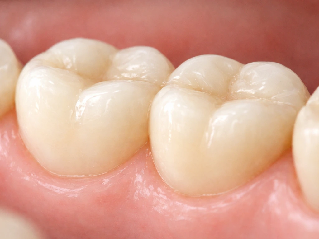
The 'double tooth' scenario, where a tooth looks like two crowns merged into one, is a developmental anomaly. It happens during the embryonic stage of tooth formation, usually in the first trimester of pregnancy, when tooth germs (the tiny cellular blueprints of future teeth) either collide and merge or attempt an incomplete split. Once the teeth have erupted, this kind of joining is already baked in and permanent.
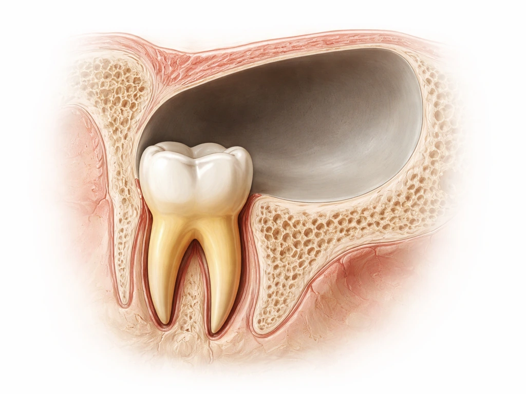
The 'locked tooth' scenario is different. Here, an already-formed tooth fuses to the surrounding jawbone because the membrane separating the tooth root from the bone breaks down. The tooth doesn't merge with another tooth; it merges with bone. This is ankylosis, and it most commonly shows up in baby molars that should have fallen out but instead stay stubbornly in place while the permanent teeth try to come in underneath or around them.
Tooth fusion vs gemination vs ankylosis: the key differences
These three conditions get lumped together constantly, even by patients who have already seen a dentist. Here's how to actually tell them apart.
Tooth fusion
Fusion happens when two completely separate tooth germs physically unite during development and form a single enlarged tooth. The key diagnostic clue is tooth count: if you count all the teeth in that arch and you're one short of what's expected, fusion is likely. The joined structure typically has a groove or notch on the biting surface where the two teeth attempted to remain separate. The pulp chambers inside may be joined or distinct depending on when in development the fusion occurred.
Tooth gemination
Gemination is the opposite process: a single tooth germ tries to split into two teeth but doesn't quite succeed. The result is one tooth root with two crowns, or one crown with a very prominent midline groove giving a 'twinning' appearance.
The critical difference from fusion is that tooth count is usually normal because you started with one germ and still have one root unit. Clinicians often differentiate fusion from gemination by tooth counting and pulp anatomy: fusion typically results in one tooth less than normal, while gemination typically preserves the normal number of teeth in the arch [tooth count is usually normal](https://www. ncbi. nlm.
nih. gov/books/NBK574555/). Dentists distinguish the two partly through X-rays looking at pulp anatomy and partly by counting teeth. In everyday clinical practice, the distinction matters because it affects the treatment plan.
Tooth ankylosis
Ankylosis is not a developmental double-tooth situation at all. It happens after a tooth has formed and sometimes after it has erupted, when the periodontal ligament (the thin shock-absorbing membrane between the root and the jawbone) is damaged and bone grows in to fill the space. The tooth fuses solidly to the bone. The most visible sign is infraocclusion: the ankylosed tooth sits lower than its neighbors because the jaw keeps growing around it while the fused tooth stays frozen in place. Primary molars are the most common victims, with studies showing an incidence of roughly 8 to 14 percent in baby teeth.
A quick note on concrescence
There's a fourth, less-discussed condition worth knowing about: concrescence. This is where the roots of two adjacent teeth are joined by cementum (the hard outer layer of the root) without the inner dentin or crowns being involved. It can happen developmentally or as a result of chronic inflammation. The teeth look normal from the outside, but extraction becomes extremely complicated because pulling one risks the other. Concrescence is often only discovered on an X-ray taken for another reason.
| Condition | How it forms | Tooth count effect | What joins | Most common location |
|---|---|---|---|---|
| Fusion | Two tooth germs unite during development | One fewer tooth than normal | Crowns and often roots (dentin + pulp) | Front teeth, primary dentition |
| Gemination | One tooth germ attempts to split | Normal tooth count | Crown divided, shared root | Upper front teeth |
| Ankylosis | PDL destroyed, bone fills the space | Normal tooth count | Root fused to jawbone | Primary molars |
| Concrescence | Cementum overgrowth joins adjacent roots | Normal tooth count | Roots joined by cementum only | Upper molars, adults |
Common causes and risk factors
Fusion and gemination are developmental anomalies, meaning they originate during tooth germ formation, typically in the early weeks of fetal development. The exact cause is not always identifiable, but the leading factors include genetics, physical pressure between adjacent tooth germs, and developmental disturbances affecting the dental lamina (the tissue that gives rise to all teeth). If a parent had a fused or geminated tooth, there is a higher chance their child will too. Some cases are associated with syndromes affecting facial development, but many cases appear in otherwise healthy children with no family history.
Ankylosis has a different set of triggers. In children, it most commonly follows trauma to a primary tooth: a knock, fall, or impact that damages the periodontal ligament. Infection around a tooth root can also destroy the PDL. Once that membrane is compromised, the body treats the gap as an injury site and fills it with bone, locking the tooth in place. Occasionally ankylosis in permanent teeth follows replantation (when a knocked-out tooth is put back in the socket) or significant orthodontic trauma.
- Family history of fused or double teeth (genetic predisposition)
- Physical crowding or pressure between adjacent tooth germs in utero
- Trauma to baby teeth, especially impacts that damage the periodontal ligament
- Chronic periapical infection destroying the PDL over time
- Tooth replantation after avulsion (being knocked fully out)
- Developmental syndromes affecting craniofacial structures
- Idiopathic causes (no identifiable reason, which is common)
How to recognize it: signs and symptoms to watch for
Parents are often the first to notice something is off, usually when a child's teeth don't look right during eruption. Adults occasionally discover a fused or geminated tooth in themselves after seeing dental X-rays for the first time in years. Here are the signs that should prompt a closer look.
Signs of fusion or gemination
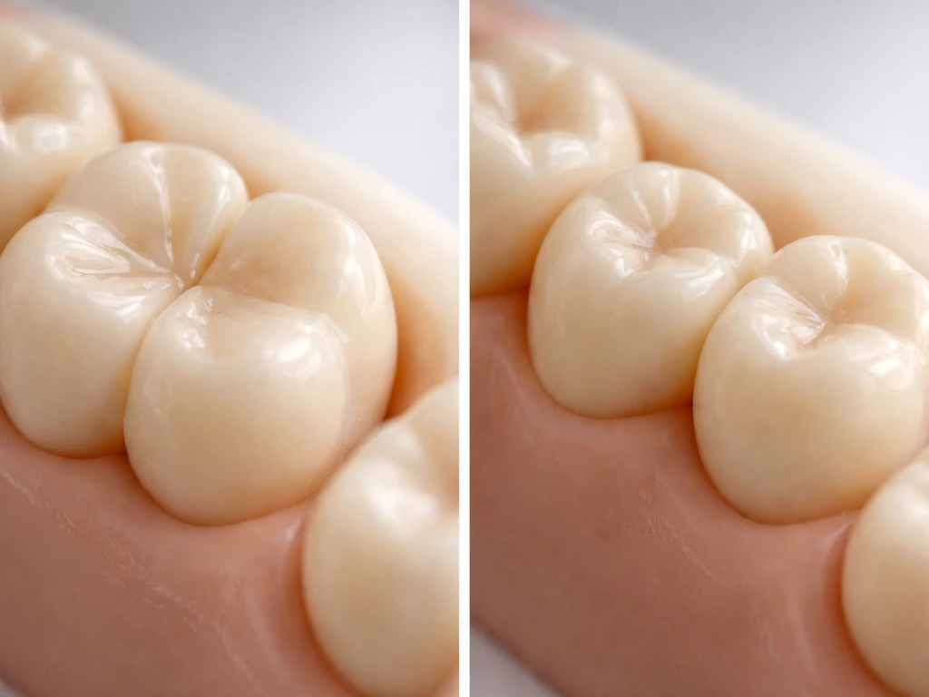
- One tooth that appears noticeably wider than its neighbors
- A visible groove, notch, or indentation running vertically down the middle of a tooth crown
- A gap or crowding issue that doesn't match the number of teeth present
- Missing tooth in the adult count when a large double-looking crown is present (suggests fusion)
- Difficulty flossing between a wide tooth and its neighbors due to the abnormal shape
- Hypersensitivity along the groove, which can trap food and harbor decay
Signs of ankylosis
- A baby tooth that has not fallen out well past the normal timeframe
- The affected tooth sits visibly lower than its neighboring teeth (infraocclusion)
- No normal tooth 'give' when you press on it (ankylosed teeth feel rigidly solid, like pressing on bone)
- A dull, flat sound when the tooth is tapped with a dental instrument, compared to the higher pitch of a healthy tooth
- Adjacent teeth tilting toward the low-sitting tooth over time
- The permanent tooth underneath erupting to the side or failing to erupt at all
One symptom that surprises people: fused or geminated teeth can develop cavities much faster than normal teeth because the groove running down the middle is nearly impossible to clean properly with a toothbrush. If you or your child has a wide tooth with a central groove and seems to get decay in that spot repeatedly, that's a strong clinical hint something structural is going on.
How dentists confirm it: exam, X-rays, and advanced imaging
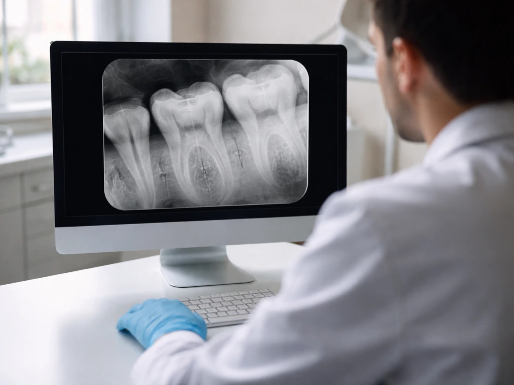
A good dentist can often make a preliminary call just from a clinical exam: counting teeth, checking crown shape, assessing mobility, and tapping teeth to listen for the tone difference. But imaging is essential to confirm what's actually happening and to plan treatment.
Standard dental X-rays
A periapical X-ray (the kind that shows the whole tooth from crown to root tip) is usually the first imaging step. It can reveal whether the pulp chambers are joined or separate (helping distinguish fusion from gemination), show the obliteration of the periodontal ligament space in ankylosis, and identify whether a permanent successor tooth is developing underneath an ankylosed primary molar. A panoramic X-ray gives a broader view of the entire dentition and is particularly useful for spotting how the anomaly is affecting tooth alignment throughout the arch.
CBCT (cone beam CT) scanning
For complex cases, especially when surgery or extraction is being considered, a cone beam CT provides a three-dimensional view that a flat X-ray simply cannot offer. CBCT is especially valuable for concrescence (where root fusion is the issue) because the exact location and extent of the joined cementum determines how risky extraction will be. It's also used when a fused or geminated tooth has roots that are curved or unusually shaped, or when an ankylosed tooth's bony union needs to be fully mapped before surgical separation is attempted.
The combination of clinical exam and appropriate imaging usually gives a definitive diagnosis. If your dentist is uncertain after a standard X-ray, asking about CBCT is entirely reasonable, particularly for a child whose permanent dentition is at risk.
Can fused or ankylosed teeth separate on their own?
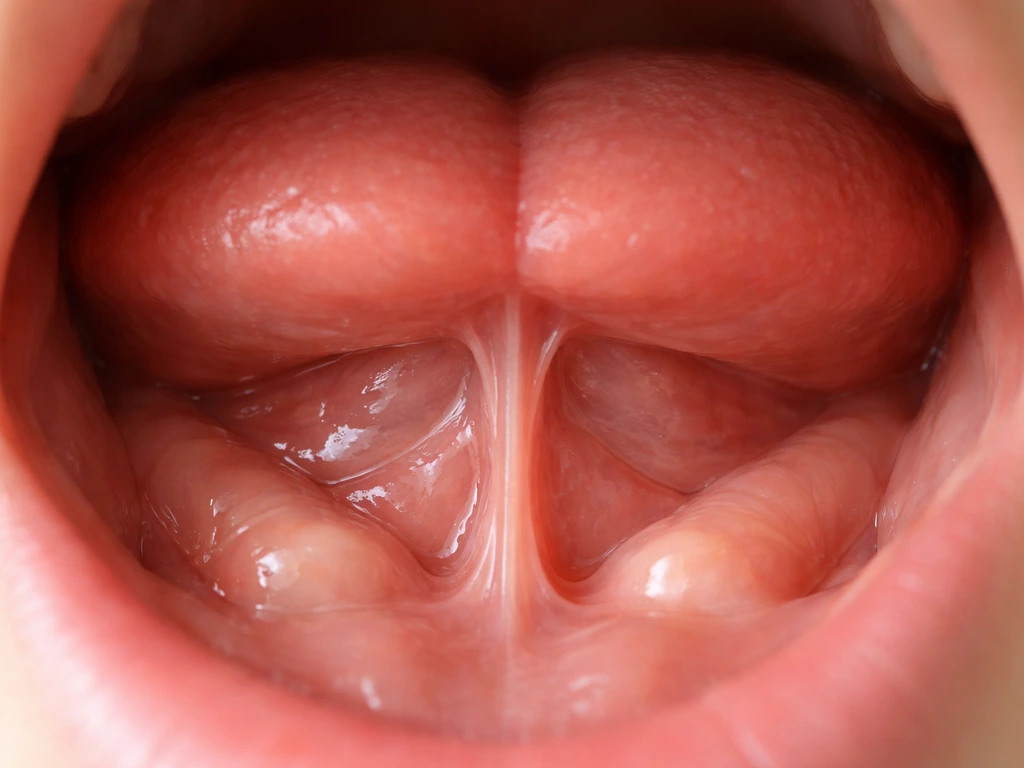
No. This is one of the most important things to understand about this topic, and it ties directly into the broader question of what teeth can and cannot regenerate or undo. Concerns like “can teeth grow under your tongue” are different from fusion, gemination, and ankylosis, and they usually point to other causes that need a proper dental or medical evaluation. Once fusion has occurred during development, the joined structure is permanent.
The teeth will not separate on their own after eruption. There is no biological mechanism for two fused tooth germs to 'un-fuse. ' The same applies to ankylosis: once bone has replaced the periodontal ligament, the body will not spontaneously re-create that membrane and free the tooth. These are one-way biological events.
This mirrors the general principle that applies across dental biology: enamel has extremely limited self-repair capacity (remineralization can fill microscopic surface damage but not structural loss), dentin has very limited capacity, and the periodontal ligament, once fully obliterated by bone, does not regenerate on its own. Teeth do not re-grow and joined dental structures do not un-join without intervention. The question is not whether the condition will resolve naturally (it won't) but whether and how treatment can improve the outcome.
There is, however, an important nuance for ankylosed primary teeth: in some mild cases, particularly when a baby molar is only slightly infraoccluded and a healthy permanent tooth is developing on schedule beneath it, monitoring without immediate intervention is a legitimate strategy. The permanent tooth's eruption force can sometimes displace a mildly ankylosed primary tooth over time. But this is not the tooth 'separating from bone spontaneously'; it's the successor tooth physically pushing the ankylosed one out.
Treatment options and when you should act now

The right treatment depends entirely on which condition you're dealing with, which teeth are involved, the patient's age, and whether the anomaly is causing functional or aesthetic problems. Here's how the decision tree typically works.
For fusion and gemination
- Monitoring and observation: If the double tooth is a primary (baby) tooth, no pain, and the permanent successor is developing normally, watching and waiting is often appropriate. The primary tooth will eventually be lost.
- Preventive sealing and restorations: The groove running down the center of a fused or geminated crown is a decay trap. Sealing it early with a dental sealant or composite resin can prevent cavities from developing in that vulnerable channel.
- Cosmetic reshaping and bonding: For erupted permanent fused or geminated teeth that are causing aesthetic concerns, composite bonding or porcelain veneers can improve appearance without separating the teeth.
- Endodontic (root canal) treatment: If the pulp becomes infected due to deep decay in the groove or structural exposure, root canal treatment may be needed. Complex pulp anatomy in fused teeth can make this technically challenging.
- Extraction and orthodontic space management: If the double tooth is creating significant crowding, bite problems, or alignment issues, extraction followed by orthodontic treatment is sometimes the best long-term solution. An orthodontist and dentist should coordinate this plan.
- Surgical separation: Physically separating two fused teeth is occasionally attempted but is technically complex, rarely successful without significant damage to both teeth, and generally reserved for specific cases where the teeth have clearly separate root systems confirmed by CBCT.
For ankylosis
- Monitoring (mild cases in young children): If the infraocclusion is minor and the permanent tooth is on schedule, periodic monitoring with X-rays every 6 to 12 months is reasonable.
- Extraction of the ankylosed primary tooth: This is often the definitive treatment when the infraocclusion is progressing, the permanent tooth is being blocked, or adjacent teeth are tipping. Extraction of an ankylosed tooth can be more difficult than a standard extraction because the root is fused to bone, sometimes requiring surgical removal.
- Bone grafting after extraction: Once an ankylosed molar is removed, the site may have a bone defect that benefits from grafting before or alongside orthodontic or implant treatment.
- Orthodontic treatment: After the ankylosed tooth is removed, orthodontics may be needed to correct the tipping of adjacent teeth and create proper space for the permanent successor.
- Implant or prosthetic options: If the permanent successor never developed or erupted in a poor position due to the ankylosis, an implant or bridge may eventually be the best way to restore the space.
When to act right now
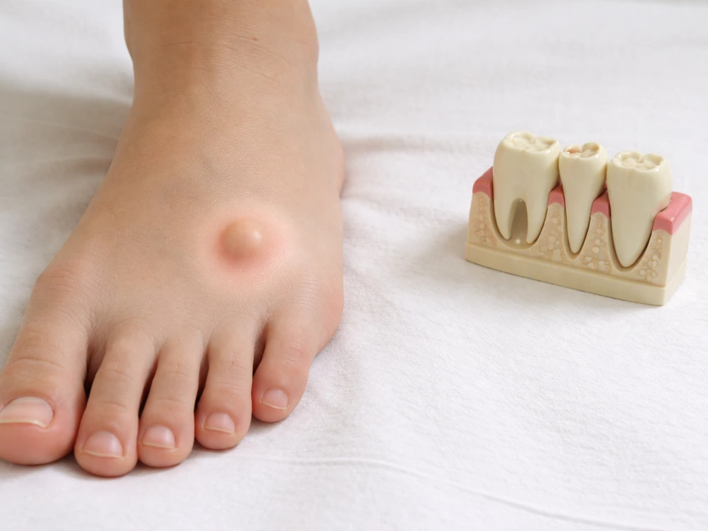
Don't wait if any of the following apply: a child's baby tooth is sitting noticeably below the gum line while permanent teeth are erupting around it; an adult tooth is failing to erupt in its expected position; you or your child has a wide 'double' tooth with a groove that already has visible decay or sensitivity; or a dentist has mentioned infraocclusion but hasn't explained the implications. Some people also wonder whether teeth can grow on your feet, but dental tooth conditions are specific to the mouth and jaw a child's baby tooth is sitting noticeably below the gum line. These are situations where delayed treatment can lead to permanent tooth loss, significant crowding, or bone loss that becomes progressively harder to correct. The earlier ankylosis in a primary molar is identified and managed, the more options remain available.
If you're unsure whether what you're seeing is fusion, gemination, ankylosis, or something else entirely (like a retained baby tooth, an impacted tooth, or even an unusual shape variant), the right move is a clinical exam with dental X-rays. A general dentist can usually make the initial diagnosis and refer you to an oral surgeon, orthodontist, or pediatric dentist depending on what's found. This is not a 'wait and see if it gets worse' situation; it's a 'get the imaging and understand what you're dealing with' situation. The earlier you have a clear diagnosis, the better the treatment options.
One final note: the internet is full of posts from people wondering if an unusual-looking tooth is something sinister. In the vast majority of cases, a fused or geminated tooth in a child is a manageable developmental variant, not an emergency and not a sign of anything dangerous. Sometimes, dental infections or abnormal root positioning can also raise concerns about whether a tooth root could affect the sinus area root growth into the sinus. But it does need to be on your dentist's radar, tracked over time, and treated proactively before it creates downstream problems for the permanent dentition. Get it documented, get a plan, and revisit it at every checkup.
FAQ
If two teeth look joined, can a dentist separate them after they erupt?
It depends on whether the joined appearance is due to fusion/gemination (tooth germ development) or ankylosis (bone fused to the tooth). With fusion or gemination, you typically cannot separate the structure after eruption. With ankylosis of a primary tooth, dentists may monitor mild cases because the permanent successor can sometimes push the primary tooth out, but that is not the same as “reversing” the fusion.
My child keeps getting cavities in the groove of a “double-looking” tooth, does that confirm fusion or gemination?
Not always. A wide tooth with a groove often points to fusion or gemination, but repeated decay can also come from plaque retention, developmental enamel defects, or an incorrect bite that traps food. The safest step is to get X-rays to confirm pulp and root anatomy before assuming it is a joined-tooth condition.
If a tooth feels stuck, does that automatically mean it is ankylosed?
You generally should avoid trying to “test” or move the tooth at home. Ankylosis can make a tooth feel immobile, but so can other issues like retained deciduous roots, crowding, or an impacted/abnormally erupting tooth. Only an exam plus imaging can tell whether the immobility is from bone fusion.
How soon should you see a dentist for ankylosis in a primary tooth?
Yes, but the treatment urgency differs. Ankylosed primary molars can affect space and eruption timing of the permanent teeth, so dentists often set a follow-up schedule rather than waiting for it to get worse. If there is infraocclusion with abnormal spacing, early documentation and a plan tend to preserve more options.
When is cone beam CT necessary for suspected fusion, gemination, or concrescence?
CBCT is usually reserved for cases where 2D imaging is not enough for surgical planning or when root complexity makes the risk hard to assess. Common reasons include suspected concrescence, unusual or curved roots, or when an extraction separation would be risky. Dentists weigh the added detail against the additional radiation and the clinical need.
What can we do at home to prevent cavities on a fused or geminated tooth groove?
If a fused or geminated tooth has a persistent central groove, flossing and targeted cleaning are important, but toothbrush alone often does not reach the groove well. Many dentists recommend special brushes or gentle interdental cleaning tools and may apply sealants or fluoride strategies to reduce decay risk. The exact plan depends on where the groove and margins are and how deep it is.
Is a panoramic X-ray enough to diagnose fusion versus gemination versus ankylosis?
A panoramic X-ray is helpful for seeing the overall dentition and eruption patterns, but it cannot replace a periapical image for determining pulp chamber relationships or the periodontal ligament space detail. Dentists commonly use a periapical film first, then add panoramic or CBCT depending on what they find.
What signs mean we should not delay getting imaging and a diagnosis?
If a child has a wide, grooved tooth, repeated “same spot” decay, or a baby tooth that does not fall out while permanent teeth are erupting, that combination is worth prompt evaluation. It does not mean an emergency, but delaying the diagnosis can reduce orthodontic and space management options later.
Could an ankylosed baby molar cause long-term problems, and is space maintenance ever needed?
If a primary tooth is ankylosed or does not exfoliate on time, a dentist may discuss space maintenance or orthodontic monitoring, depending on what the permanent successor looks like on X-ray. The goal is to prevent crowding or abnormal eruption paths, which can become harder to correct once space is lost.
How would concrescence change the approach compared with fusion or gemination?
Concrescence can look subtle externally, and the key issue is that extraction can be hazardous because cementum may join roots in a way that is not obvious in a quick exam. That is why dentists sometimes request a 3D scan when extraction is planned but the roots appear unusually connected or extraction risk is unclear.
Next Articles
Can Teeth Grow Under Your Tongue? Causes and What to Do
Can teeth grow under your tongue? Learn causes of bumps, real tooth limits, and next steps for urgent evaluation.
Can a Tooth Root Grow Into the Sinus? What to Know
Can an upper tooth root grow into the sinus? Learn anatomy, X-ray clues, symptoms, tests, risks, and treatment options.
Can Teeth Grow on Your Feet? Causes and What to Do
No, teeth can’t grow on feet. Learn causes of tooth-like bumps, red flags, and next steps for diagnosis.