No, a tooth cannot simply grow under your tongue the way teeth erupt through your gums. The floor of your mouth is not dental tissue, and there is no tooth bud sitting there waiting to emerge. If you are feeling a bump, lump, or hard spot under your tongue, the overwhelming likelihood is that it is a salivary gland issue, a benign cyst, a minor infection, or in rare cases a developmental anomaly involving a misplaced (ectopic) tooth. Any of those things need attention, but none of them are your body spontaneously regrowing a tooth in a new location.
Can Teeth Grow Under Your Tongue? Causes and What to Do

Marcus Holloway
13 Jun 2026
What is actually under your tongue (a quick anatomy check)
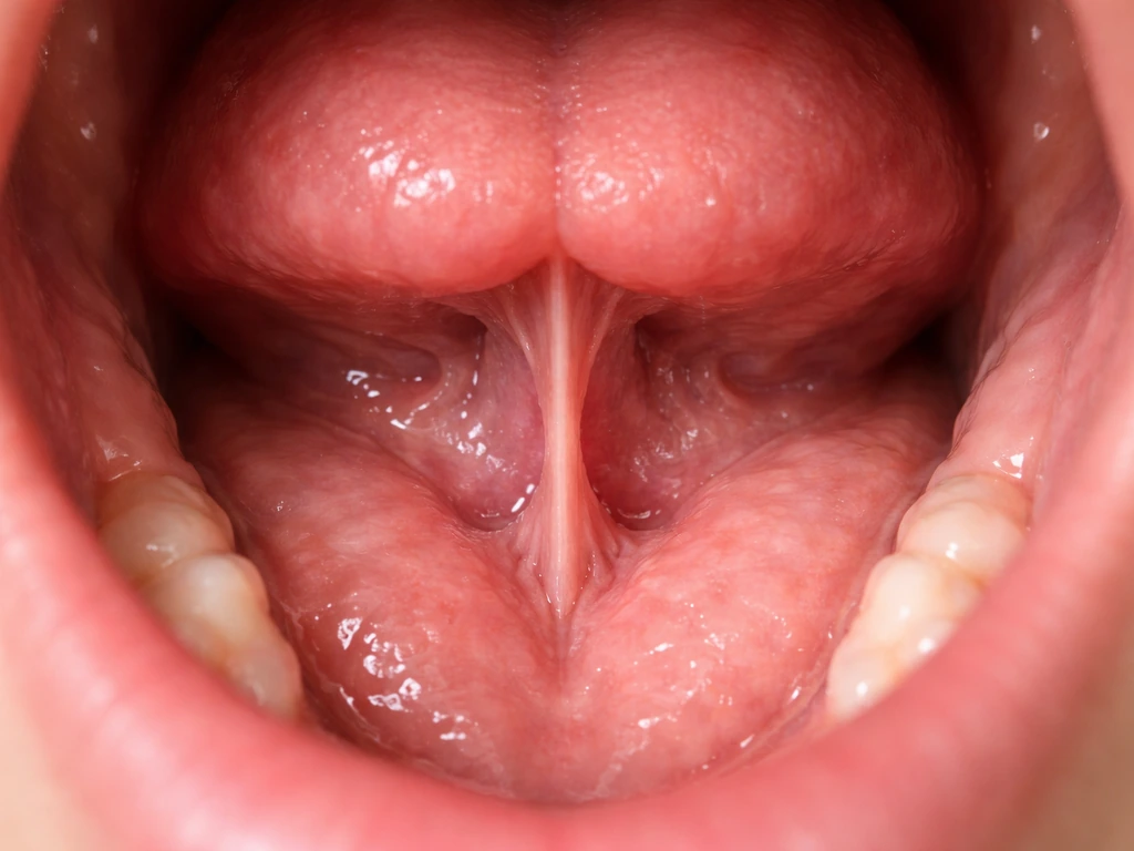
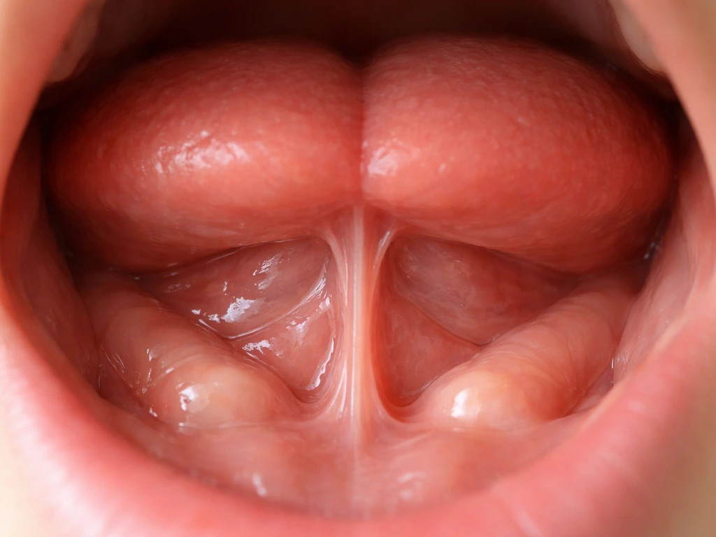
The area beneath your tongue is called the floor of the mouth. It is packed with soft-tissue structures, none of which are teeth. The lingual frenum is the thin band of tissue connecting the underside of your tongue to the floor of the mouth. On either side of its base are two small raised bumps called sublingual papillae (sometimes called the sublingual caruncles), and these are actually the openings of Wharton's duct, which drains saliva from your submandibular glands.
Just beneath the mucosa on each side sit the sublingual glands themselves, tucked above the mylohyoid muscle and below the sublingual fold. That is a lot of plumbing for a small space, and any one of those structures can swell, get blocked, or form a cyst. There is zero dental anatomy down there under normal circumstances. No tooth buds.
No enamel-forming cells. No roots. So when something unexpected shows up, you are dealing with soft-tissue pathology, not dentistry in the traditional sense.
Common reasons you might feel something tooth-like under your tongue
Several conditions can produce a hard, firm, or lumpy feeling under the tongue that people sometimes describe as feeling like a tooth pushing through. Here is what is actually behind most of those cases.
Salivary stones (sialolithiasis)
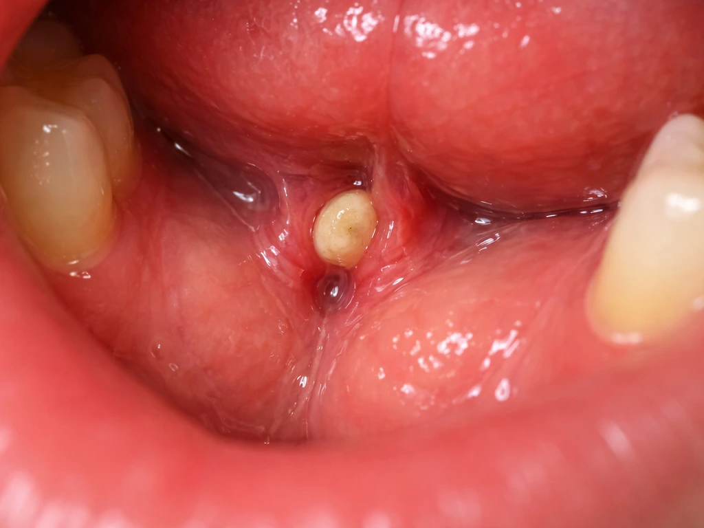
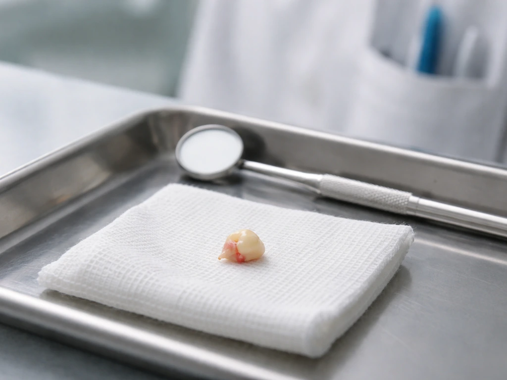
This is probably the most common culprit for something that feels hard and tooth-like under the tongue. About 80% of salivary stones form in the submandibular glands and block Wharton's duct, which opens right at the base of your lingual frenum. When a stone blocks that duct, saliva backs up, the gland swells, and the area under your tongue can become noticeably firm or painful.
The pain typically gets worse when you eat, because eating triggers your salivary glands to produce more saliva, and that saliva has nowhere to go. A stone sitting near the duct opening can sometimes be felt directly with a fingertip. It is calcified, dense, and absolutely can feel like a small piece of tooth. Around 70 to 80 percent of submandibular stones are dense enough to show up on a basic X-ray.
Ranula (a salivary cyst)
A ranula is a type of pseudocyst that forms when saliva leaks out of the sublingual gland and pools in the surrounding tissue. It usually presents as a painless, soft swelling on one side of the floor of the mouth. Superficial ranulas often have a bluish or translucent color that gives them a somewhat distinctive look, but deeper ones can appear pinkish and are easy to confuse with other lesions. A ranula is not hard like a tooth, but a large one can create enough bulk under the tongue that it feels like something structural is growing there.
Mucocele
Mucoceles are similar to ranulas but typically smaller and more commonly caused by trauma (like biting your cheek or lip). They can occasionally form on the floor of the mouth. Superficial ones look bluish and translucent. Deeper ones look more like regular pink tissue. They are generally not painful and tend to come and go.
Salivary gland infection (sialadenitis)
If the submandibular gland gets infected, either because a stone has blocked it long enough or because of bacterial or viral causes, the resulting swelling can be dramatic. Viral sialadenitis (like mumps) can come with fever and general illness. Bacterial infections can cause the gland to swell hard and become quite tender under the jaw and under the tongue. The important thing here is that a spreading infection in this area is not something to sit on, because in serious cases it can spread to the neck and threaten the airway.
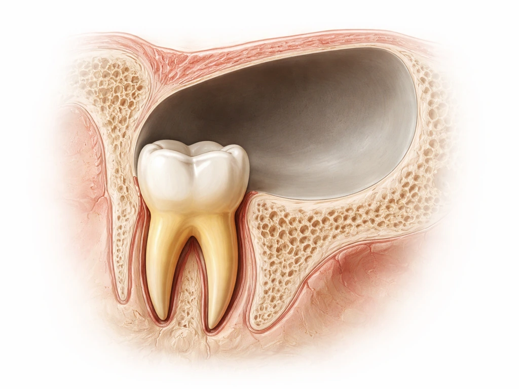
Ectopic or supernumerary tooth
This is the rare case where an actual tooth-like structure is genuinely involved, but it is still not your body spontaneously growing a new tooth. Ectopic teeth are teeth that erupt in an abnormal location due to a developmental anomaly. Supernumerary teeth are extra teeth beyond the normal 32, and they can occasionally become impacted or erupt in unexpected spots. The case report describes complications of supernumerary teeth such as impaction with [delayed eruption](https://pmc.
ncbi. nlm. nih. gov/articles/PMC12369741/) and ectopic eruption, supporting that tooth-like bumps can reflect extra teeth rather than salivary pathology.
There are documented cases of teeth erupting in places like the nasal cavity. An ectopic tooth under the floor of the mouth is exceptionally uncommon, but it is a real category of dental anomaly. An ectopic tooth can rarely erupt in other regions too, such as the nasal cavity or even near the sinus, but it would still be identified with imaging rather than being natural growth tooth erupting in the sinus.
This would require imaging to confirm and would need management by an oral surgeon.
Other possibilities
- Torus mandibularis: bony growths that appear along the inner lower jaw, sometimes near the base of the tongue area, that are completely harmless but can feel alarming
- Fibroma: a smooth, firm overgrowth of fibrous tissue, often from chronic irritation
- Dermoid cyst: a developmental cyst that can form in the floor of the mouth, sometimes filled with keratin or even hair follicle tissue
- Oral cancer: a persistent ulcer or mass on the floor of the mouth that does not heal is a warning sign that needs professional evaluation right away
What can actually grow or regenerate in this area
Let's clear up the regeneration question directly. Enamel, once lost anywhere in the mouth, cannot grow back. The Cleveland Clinic puts it plainly: enamel cannot regrow. The cells that form enamel (ameloblasts) are lost when a tooth finishes developing.
There is no mechanism in the floor of the mouth to produce new enamel, dentin, or a tooth crown. What can regenerate is oral mucosa, meaning the soft pink lining of your mouth is quite good at healing, and salivary gland tissue can recover after infection or obstruction is resolved.
There is also a specialized dental procedure called regenerative endodontics, used in necrotic immature permanent teeth in children, where the pulp-dentin complex can be biologically regenerated to encourage continued root development. But that is a clinical treatment for a specific scenario, not something that happens spontaneously, and it definitely does not produce a new tooth crown growing out of your floor of mouth. The biology simply does not work that way in adults.
A real tooth appearing under your tongue would be a true developmental anomaly, not regeneration.
Normal vs. red flag: what the symptoms are telling you
Most bumps under the tongue are benign and manageable, but some symptoms should move you toward same-day or urgent care rather than a routine appointment.
| Symptom | What it suggests | Urgency |
|---|---|---|
| Painless blue or translucent swelling on one side | Ranula or mucocele | Routine dental visit within a week or two |
| Hard lump that hurts more when eating | Salivary stone blocking Wharton's duct | Dental or ENT visit soon, within a few days |
| Swelling with fever, general illness | Salivary gland infection (sialadenitis) | See a doctor promptly, same day if possible |
| Rapid swelling, difficulty swallowing, muffled voice, drooling | Spreading infection, possible airway risk | Emergency room immediately |
| Persistent ulcer or firm lump that doesn't change over 2 weeks | Needs evaluation to rule out malignancy | Dental or medical appointment within days, not weeks |
| Firm bony ridge along the inner jaw near the tongue | Torus mandibularis (benign bone growth) | Mention at your next routine dental visit |
| Hard nodule that feels like a small pebble at the duct opening | Salivary stone near the surface | Dental or ENT evaluation within a few days |
The airway warning deserves extra emphasis. If the swelling under your tongue is progressing fast, if you are drooling because swallowing is painful, if your voice sounds muffled like you have a hot potato in your mouth, or if you are having any trouble breathing, do not wait. Go to an emergency room. A spreading infection in the floor of the mouth can compromise your airway and becomes a medical emergency quickly. Cleveland Clinic also warns that sialadenitis can become a medical emergency when swelling spreads to neck tissues and blocks the airway spreading infection in the floor of the mouth threatens the airway.
What to do today: self-check and getting assessed

Quick self-check you can do right now
- Tilt your head back in good light (a flashlight or phone torch helps). Lift your tongue and look at the floor of your mouth for any visible swelling, color change, or asymmetry between the left and right sides.
- Gently press the tip of one finger along the floor of the mouth from back to front on each side. Note whether anything feels hard, pebble-like, or tender.
- Try eating a small, sour food like a lemon wedge or a sour candy. If the swelling increases noticeably or becomes suddenly more painful within a minute or two, that is a classic sign of a blocked salivary duct and supports a salivary stone diagnosis.
- Check for fever using a thermometer. A fever alongside mouth swelling shifts this toward infection territory.
- Note how long the bump has been there and whether it changes size or fluctuates. Salivary swellings often vary with meals. A lump that stays the same size regardless of eating is more likely to be something structural.
Who to see and what to expect
For most under-tongue lumps, your regular dentist is a perfectly appropriate first stop. They will do a visual and tactile examination of the floor of the mouth (pressing along the duct path to feel for stones), check your lymph nodes, and decide whether imaging is needed. If a salivary stone is suspected, they may refer you to an oral surgeon or an ear, nose, and throat (ENT) specialist.
If the lump looks suspicious for a lesion that might need a biopsy, an oral surgeon or oral medicine specialist would handle that. Bring a timeline of when you first noticed the bump, whether it changes with meals, any pain levels, and whether you have had fever or difficulty swallowing. That information cuts the diagnostic time considerably.
Imaging and diagnostic tools your provider might use
- Dental X-ray: useful as a quick first step because 70 to 80% of submandibular stones are radiopaque (dense enough to show up on film)
- Ultrasound: good for detecting stones and assessing gland swelling without radiation, and can be done in-office
- CT scan: more definitive for stones, swelling extent, or if a spreading infection is suspected
- MRI: helpful for soft tissue lesions, ranulas extending into the neck, or when more detail on a mass is needed
- Sialoendoscopy: a small camera threaded into the salivary duct to directly visualize and sometimes remove stones, used by oral surgeons and ENT specialists
- Sialography: injection of contrast dye into the duct with X-ray, less commonly used now but still employed in some settings
Treatment options depending on what you are actually dealing with
Salivary stones
Small stones near the duct opening can sometimes be expressed on their own with sialagogues (things that stimulate saliva flow, like lemon juice or sour candy) combined with gentle massage over the gland. A clinician can sometimes manually manipulate or probe the duct to dislodge a small stone.
For larger or more stubborn stones, the main modern options are sialoendoscopy (where the stone is retrieved through the duct using a small instrument) or surgical removal, sometimes with removal of the gland itself in recurrent cases. Sialoendoscopy has a reported calculus removal success rate of around 88%, while surgical removal of the gland is essentially 100% effective at preventing recurrence. Extracorporeal shock-wave lithotripsy (using targeted sound waves to break up stones) is also used in some settings.
If infection has developed alongside the obstruction, antibiotics will be part of the treatment plan.
Ranula
A small, asymptomatic ranula might be watched rather than immediately treated. For symptomatic or enlarging ranulas, the most effective treatment is complete excision of the ranula together with the affected sublingual gland. The data on recurrence is stark: marsupialization alone (opening and draining the cyst without removing the gland) has recurrence rates reported between 61 and 89%. Removing just the ranula without the gland produces recurrence rates of 57 to 69%. Complete excision of the ranula plus the sublingual gland drops recurrence to about 0 to 2%. For a ranula that has extended into the neck (called a plunging ranula), imaging with CT or MRI is used first to map the extent before surgery.
Salivary gland infection
Bacterial sialadenitis is treated with antibiotics, hydration, warm compresses, salivary massage, and sialagogues to keep saliva flowing. Viral causes like mumps are managed supportively. If an abscess forms, it needs drainage. Spreading infections that involve the deep neck spaces require hospitalization and IV antibiotics; airway protection becomes the priority.
Ectopic or supernumerary tooth
If imaging confirms an actual ectopic or supernumerary tooth in the floor of the mouth, an oral surgeon handles removal. The approach depends on the tooth's position, size, and relationship to surrounding structures like nerves and the duct. This is not an emergency unless the tooth is causing significant symptoms, but it does need to be addressed.
Benign structural growths (torus, fibroma, dermoid cyst)
Tori that are small and not causing problems are usually left alone. Larger ones that interfere with speaking, eating, or denture fit can be surgically reduced. Fibromas and dermoid cysts are surgically excised if they are symptomatic or growing. Recurrence of fibromas is low when the source of irritation is also addressed.
Persistent lesion or ulcer
Any lesion on the floor of the mouth that has not resolved in two weeks needs to be evaluated by a dentist or oral medicine specialist. The ADA recommends visual and tactile examination and follow-up to determine whether biopsy or further evaluation is needed. The floor of the mouth is a high-risk site for oral cancer. Do not self-diagnose this one, and do not wait months hoping it resolves on its own.
Prevention and long-term care (what actually helps)
Because the most common under-tongue issues relate to the salivary gland system, most prevention advice centers on keeping that system working well. Dehydration is a major contributor to salivary stone formation because it makes saliva thicker and more prone to mineral deposits. Drinking adequate water throughout the day is probably the single most practical preventive step. Stimulating regular saliva flow with meals, staying away from extremely low-moisture diets for extended periods, and avoiding smoking (which affects saliva composition) all contribute to a healthier salivary environment.
For the dental side of things: there is no intervention that regenerates tooth structure that has already been lost in the floor of the mouth or anywhere else in an adult. Enamel cannot regrow. Fluoride helps remineralize early enamel damage before it progresses to a full cavity, but it is not regrowing structure, it is reinforcing what remains. Keeping your regular dental appointments matters because your dentist does a floor-of-mouth examination as part of routine care and is in a position to catch something early, long before it becomes a problem you can feel yourself.
Parents of young children should know that unusual lumps in a child's floor of mouth should always be checked, especially if a child is still in mixed dentition (the phase of losing baby teeth and gaining permanent ones). Ectopic eruption and supernumerary teeth are more likely to be caught in childhood, and early intervention usually means simpler treatment. For adults and seniors, the calcification risk for salivary stones increases with age, and any new hard lump under the tongue in a person over 40 deserves prompt evaluation rather than watchful waiting at home.
One final myth worth addressing: the internet occasionally suggests that certain supplements or protocols can stimulate new tooth growth. There is no evidence for this in humans. Sharks regrow teeth. Humans get two sets and that is the end of the line biologically, at least for now.
The same logic applies to the floor of the mouth: the tissue there is not capable of producing new dental structures without a significant developmental anomaly. If you are ever curious how this compares to other unusual tooth-related questions, things like whether teeth can appear in completely non-oral locations follow the same logic of rare developmental anomaly rather than natural regrowth, and those cases are almost always identified through imaging rather than a bump you can feel at home.
If you are wondering whether teeth can grow on your feet, that is not something that happens naturally either.
FAQ
If the bump under my tongue comes and goes, does that make it less likely to be something serious?
Not usually. A transient “hard spot” after a meal is more consistent with a salivary duct blockage or irritation (for example, a small stone near Wharton’s duct) rather than any dental growth. If the lump persists beyond about two weeks, enlarges, or becomes painful, it should be examined.
How can I tell whether it’s a salivary gland issue versus a tooth problem?
Yes, because salivary gland problems can mimic dental pain. Stones and sialadenitis often feel like a tooth is coming in, but the pattern is usually meal-related (worse when salivating) and may come with swelling near the duct opening rather than a gum eruption site.
Is it safe to try to push the bump or drain it at home?
You should not try to “pop,” cut, or squeeze it. That can worsen infection or cause the swelling to spread in the floor of the mouth. If you suspect a stone, gentle hydration, warm compresses, and sialagogues can help, but any procedure to force it out should be done by a clinician.
What tests does a dentist or ENT typically use to find out what the lump is?
In many cases, dental or medical imaging is what distinguishes an ectopic tooth from other lesions. For suspected salivary stones, basic X-ray may show dense stones, while soft-tissue concerns, deeper ranulas, or uncertain masses may require ultrasound, CT, or MRI.
When should I go to urgent care versus scheduling a routine dental visit?
It depends on severity, not just your age. If you have rapidly increasing swelling, drooling, muffled voice, trouble swallowing, or any breathing difficulty, seek emergency care immediately. Otherwise, a new lump that has not resolved within two weeks should be scheduled promptly, especially if you are over 40.
What symptoms mean the bump could be an infection and not just a blockage?
If you have fever, escalating pain, pus-like drainage, or feel generally unwell along with a swelling under the tongue or under the jaw, treat it as potentially infectious. Those situations often require prescription treatment and sometimes urgent evaluation to rule out an abscess or deeper neck involvement.
Does the color or side of the swelling help identify ranula versus a salivary stone?
A ranula is commonly described as bluish or translucent when superficial, and it is often on one side of the floor of the mouth. It tends to be more soft or fluctuant than a true hard “tooth-like” structure, but deeper ranulas can look less obvious and feel bulkier.
Can I use lemon drops or other sour foods if I suspect a salivary stone?
Sialagogues (sour candy or lemon) and hydration are helpful when there is suspected duct blockage, but they can also aggravate pain if infection is already present. If you feel worse quickly with food, have fever, or the swelling is rapidly expanding, stop home measures and get evaluated.
If it keeps happening, what are the options beyond “wait and see”?
Yes, especially if you have recurring episodes. If stones or swelling keep coming back, clinicians may discuss procedures such as sialoendoscopy or, in selected recurrent cases, removal of the involved gland to reduce repeated blockage.
What are the most common non-tooth causes of a tooth-like bump under the tongue?
Common adult “false alarms” include mucoceles, ranulas, enlarged salivary glands, benign fibrous tissue, and dermoid or other cystic lesions. True ectopic or supernumerary teeth in the floor of the mouth are exceptionally uncommon, so persistent or changing lesions should still be evaluated, but don’t assume it is a tooth.
How concerned should I be about oral cancer if a lesion is on the floor of the mouth?
You should still be checked if the lump is new, growing, hard, or associated with pain or ulceration. Cancer risk is a reason not to delay evaluation, especially if it does not improve within two weeks, even though many lesions are benign.
Could my medications or dehydration be causing the problem under my tongue?
If you are taking medications that can reduce saliva (for example, some antihistamines, antidepressants, and blood pressure or bladder drugs), dryness can increase stone risk. Consider asking your clinician whether your medication list could be contributing and whether saliva-stimulating strategies are appropriate for you.
Next Articles
Can a Tooth Root Grow Into the Sinus? What to Know
Can an upper tooth root grow into the sinus? Learn anatomy, X-ray clues, symptoms, tests, risks, and treatment options.
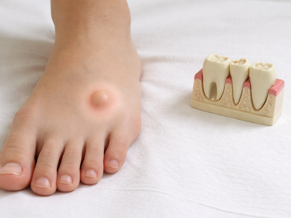
Can Teeth Grow on Your Feet? Causes and What to Do
No, teeth can’t grow on feet. Learn causes of tooth-like bumps, red flags, and next steps for diagnosis.
Do Milk Teeth Grow Back? Timelines, What to Do, Red Flags
Learn if milk teeth regrow, typical timelines for permanent teeth, what to do after loss or injury, and red flags.