A tooth root does not grow into the maxillary sinus the way a plant root grows into soil. Roots do not sprout new tissue and push through bone on their own. What actually happens is more subtle and more common than most people realize: the upper molar roots can sit so close to the sinus floor that infection, inflammation, or a difficult extraction creates a connection between your mouth and the sinus cavity. That connection, called an oroantral communication, is what causes the strange symptoms people describe. It is not root regrowth. It is anatomy plus a complication.
Can a Tooth Root Grow Into the Sinus? What to Know

Marcus Holloway
12 Jun 2026
How your upper tooth roots and the maxillary sinus are neighbors
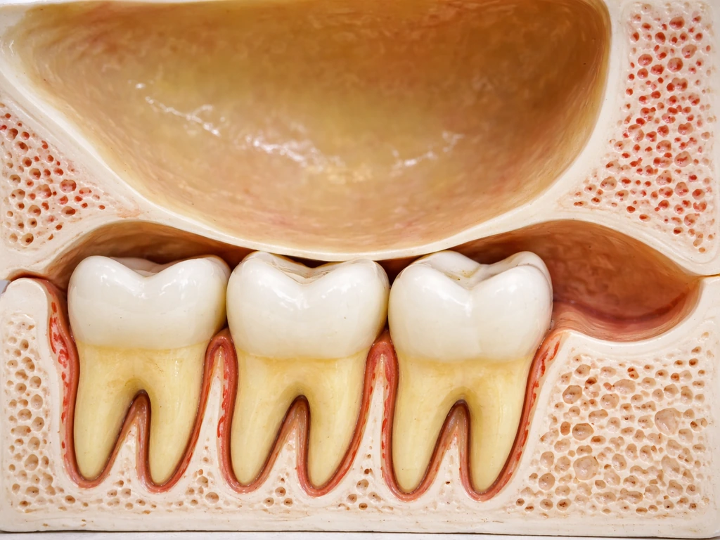
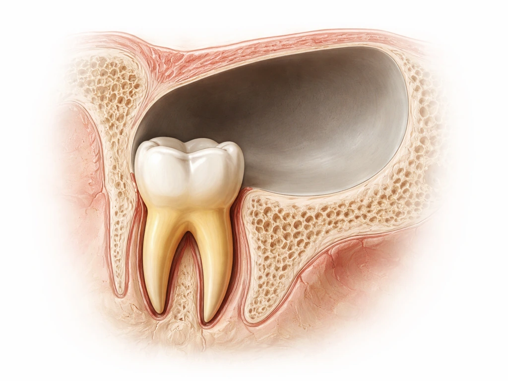
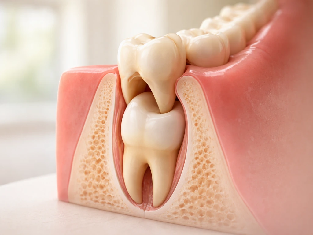
The maxillary sinus sits inside your cheekbone, one on each side, directly above the upper back teeth. The floor of that sinus is often extremely close to the roots of your upper molars and sometimes your upper premolars. CBCT imaging studies have found that in roughly half of upper molar roots, the root tip either touches the sinus floor or projects into it with no bony barrier separating them. In one measurement study, the mesiobuccal root of the upper second molar averaged only about 1.97 mm from the sinus floor. That is less than two millimeters. For some people the gap is effectively zero.
This proximity is not a defect or a disease. It is just normal anatomy that varies from person to person. Age plays a role too. Research using CBCT on thousands of roots shows a significant negative correlation between age and apex-to-sinus distance, meaning older adults tend to have roots that sit closer to the sinus floor. Wisdom tooth roots, which finish developing in young adulthood, can also end up very close to the sinus depending on how the second and third molars are positioned. The key point is that the closeness is built-in, and most people never have a single problem because of it.
So can a root actually "grow into" the sinus?
Not in the biological sense of new root tissue generating and extending through the sinus membrane. Root development follows a strict developmental window during childhood and adolescence. Once a root is fully formed, it does not keep growing. There is no mechanism in adult dental biology that would cause a mature root to spontaneously push through bone, through the sinus membrane, and into the air-filled cavity above it.
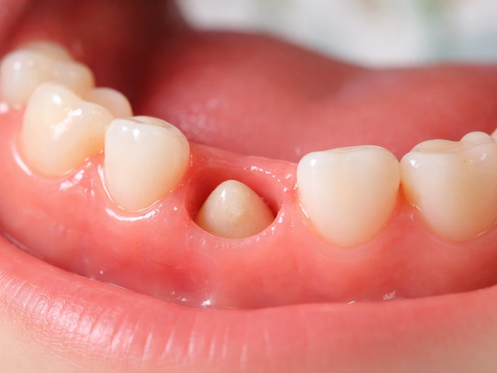
What people call a root "in the sinus" on imaging is usually one of three things. First, a root that was always anatomically close enough to indent or rest against the sinus floor, which is common and often harmless. Second, a root whose surrounding bone has been eroded by a periapical infection, cyst, or granuloma, so that the sinus membrane is now exposed to the infection pathway. Third, a root fragment that was accidentally displaced into the sinus during an extraction, which is an iatrogenic complication, not biology. The distinction matters enormously because each scenario has a completely different treatment path.
Here is something that surprises people: even when CBCT imaging shows a root appearing to protrude into the sinus, histological studies found actual sinus membrane perforation in only 14 to 28 percent of those cases. Proximity on a scan does not automatically mean the membrane is breached. Context and symptoms are everything.
Symptoms that suggest your sinus and tooth are connected

The classic symptom cluster for an oroantral communication or odontogenic sinusitis is almost always one-sided. Common symptoms reported for odontogenic maxillary sinusitis include unilateral purulent rhinorrhea and a foul smell or taste, with additional associated complaints such as post-nasal drip and cheek pain often alongside dental pain common symptoms include unilateral purulent rhinorrhea and foul smell/taste. That asymmetry is a major clue. If your regular sinus trouble is typically bilateral, and suddenly you have pressure, discharge, or congestion only on one side, especially near an upper back tooth, that warrants attention.
- Unilateral purulent nasal discharge, often with a foul smell, sometimes described as a bad taste at the back of the throat
- Pressure or pain in one cheek that feels different from typical sinus headaches
- Air passing through an extraction socket or gap when you breathe through your nose
- Liquid leaking into your nose when you drink
- A voice change or nasal resonance quality that appeared after dental work
- Persistent postnasal drip only on one side
- Swelling or discharge along the upper gum line near a back molar
- Sinus congestion that does not respond to standard nasal medications
The air-passage symptom is one of the most telling. If you can feel or hear air moving through an extraction socket when you exhale through your nose, that is a direct sign of an oroantral communication. You should not try to force this or test it repeatedly. Just note it and call your dentist or oral surgeon.
How dentists diagnose a sinus-tooth connection
The clinical exam
A dentist or oral surgeon will first look at the socket or area of concern directly. The Valsalva maneuver is a standard clinical test: you are asked to try to exhale gently through your nose while your nostrils are pinched closed. If an oroantral communication is present, air or bubbling at the socket confirms it. Worth knowing: clinicians use this test carefully because forcing air through an infected connection can push bacteria further into the sinus, so it should be done by a professional, not as a DIY test at home.
X-rays and CBCT imaging
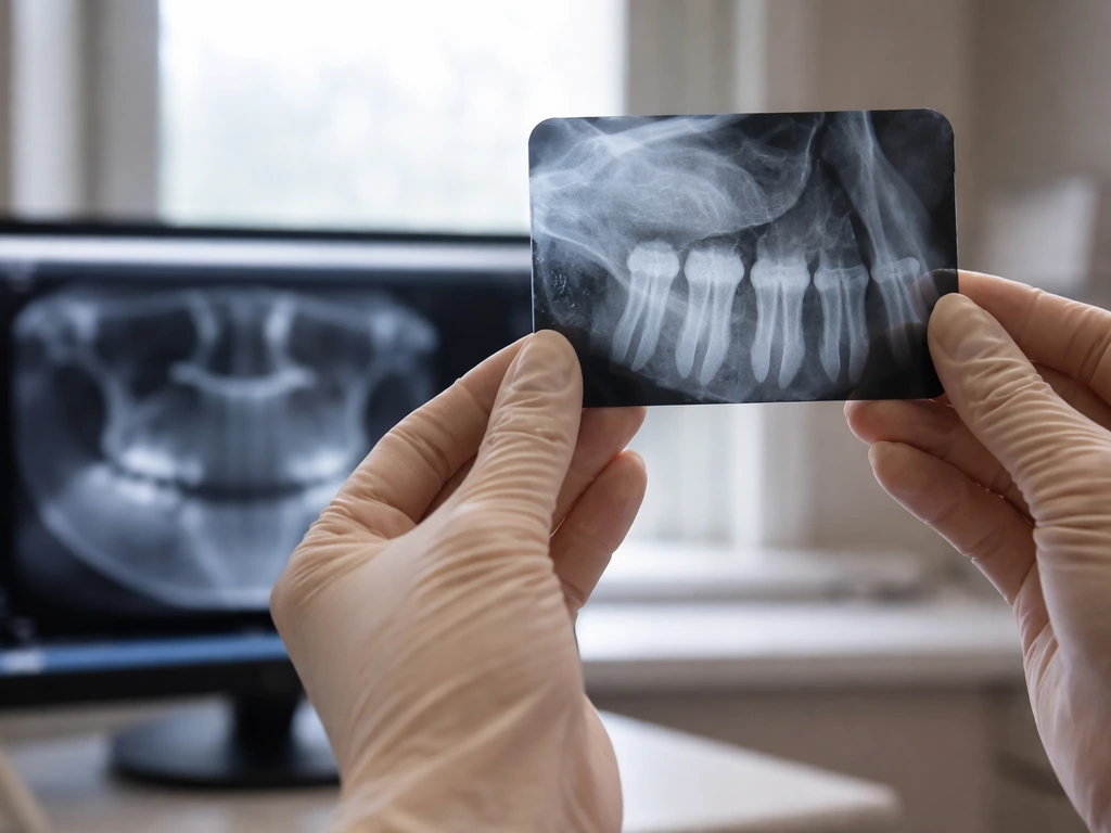
Periapical and panoramic radiographs are usually the first imaging step. They can show root proximity to the sinus floor, loss of the cortical bone line, sinus opacification, and bony defect size. A panoramic radiograph can effectively rule out a relationship if there is a clear gap visible between the root tip and sinus floor. However, when overlap or proximity is ambiguous on 2D imaging, cone beam CT (CBCT) gives a three-dimensional picture that 2D films simply cannot match. CBCT is the gold standard for seeing exactly how a root relates to the sinus, identifying sinus floor disruption, spotting displaced root fragments, detecting mucosal thickening or polyps, and planning any surgical repair. For patients whose sinus symptoms do not resolve after dental treatment, CT scanning through the sinus is also often ordered to evaluate the extent of mucosal disease before an ENT consultation.
When to treat this as urgent
Not every root-sinus proximity situation is an emergency. But certain red flags mean you should call an oral surgeon within a day or two rather than waiting for a routine appointment.
- Air or liquid passing between your mouth and nose after an extraction, persisting beyond 24 to 48 hours
- Foul-smelling or tasting discharge from an extraction socket or the nose that worsens rather than improves
- Fever combined with one-sided facial swelling and increasing pain after a dental procedure
- Unilateral sinus symptoms that started right after an extraction and are not improving after a week
- A visible opening in the socket floor after tooth removal that your dentist has not already addressed
- Any known root fragment that was not recovered after extraction, especially if sinus symptoms develop in the following weeks
The timing window matters here. Research shows that an oroantral communication can epithelialize into a chronic fistulous tract in as little as 7 to 8 days. A chronic oroantral fistula is significantly harder to close than a fresh communication, so early treatment is not optional. Similarly, odontogenic sinusitis that is left untreated can progress to chronic maxillary sinus disease that eventually requires combined dental and ENT surgery to resolve.
Treatment options, from watchful waiting to surgery
Management depends on what is actually happening, the size of any communication, how long it has been present, and whether infection is involved.
| Situation | Typical Management | Who Manages It |
|---|---|---|
| Root close to sinus, no symptoms | Monitoring, imaging follow-up, careful extraction planning if needed | General dentist or oral surgeon |
| Small OAC (<2 mm) after extraction, no infection | Clot protection, sinus precautions (no blowing nose, no straws), may heal spontaneously | General dentist or oral surgeon |
| Larger OAC (>2-3 mm) or any fistula | Surgical flap closure (buccal advancement, palatal rotation, or buccal fat pad flap) | Oral surgeon |
| Chronic oroantral fistula | Flap closure combined with endoscopic sinus surgery in most cases | Oral surgeon plus ENT |
| Odontogenic sinusitis (no OAC) | Address dental cause (extraction, root canal, retreatment) plus endoscopic sinus surgery if needed | Oral surgeon plus ENT |
| Root fragment displaced into sinus | Surgical retrieval, often endoscopically | Oral surgeon or ENT |
Sinus precautions after extraction near the sinus are important even when no communication has been confirmed. You will typically be told to avoid blowing your nose forcefully, avoid drinking through a straw, sneeze with your mouth open, and skip activities that increase sinus pressure for at least a week. These precautions protect the clot and reduce the chance of disrupting a fresh connection before it heals.
For established odontogenic sinusitis, the evidence strongly favors treating both the dental cause and the sinus together. Studies report 90 to 100 percent success rates when endoscopic sinus surgery and oroantral fistula closure are performed together. Endoscopic sinus surgery (ESS) has largely replaced the older Caldwell-Luc approach because it has fewer long-term complications. Antibiotics alone are not curative here. They may reduce acute symptoms temporarily, but without removing the dental source and restoring sinus drainage, the infection returns.
Clearing up the myths about root regrowth and sinus involvement
This is where a lot of confusion lives online. The question usually comes from a real worry: someone read that their root was "in the sinus" on their scan, or a dentist mentioned the sinus during an extraction, and they started wondering if a root somehow grew there or could keep growing. Here is what the biology actually says.
Adult tooth roots do not regenerate or extend on their own. Adult tooth roots generally do not grow together or merge; if two structures seem connected on imaging, it is usually due to proximity or a disease- or treatment-related communication can two teeth grow together. Root development is a one-time event tied to early life. There are experimental regenerative endodontic procedures being studied that can stimulate some root-like tissue in certain young patients with incompletely formed roots, but even those rarely produce perfectly normal root anatomy, and the case literature describes them as managed clinical procedures, not spontaneous growth. There is no credible scenario in which a fully-formed molar root quietly grows through the sinus floor and into the air cavity above it because of aging, diet, or anything short of extreme pathology.
What can happen over time is that infection, cyst formation, or root resorption erodes the thin bony barrier between a root tip and the sinus. This is not the root growing. It is the bone and membrane being destroyed by disease. The root stays put. The protection around it disappears. That distinction changes how you think about prevention and treatment: you are trying to catch and treat infection early, not trying to stop the root from moving.
Similarly, when people ask questions like whether teeth can grow in unusual places in the body (topics that come up across dental biology discussions) the answer always comes back to the same biological limit: tooth tissue does not self-generate and travel. In this case, proximity to the sinus is about anatomy and about what disease does to that anatomy. The root was always there. The question is whether the barrier holding everything safely apart has been compromised.
If you are dealing with unexplained one-sided sinus symptoms, a recent difficult extraction of an upper molar, persistent bad taste after dental work, or a dentist who mentioned your roots are close to the sinus, the right next step is a consultation with an oral and maxillofacial surgeon. Bring any X-rays or imaging you already have. A good exam and the right scan will answer definitively what is happening, and most of these situations have straightforward, well-established treatment once the diagnosis is clear.
FAQ
If my CBCT says a root is “into” or “in” the sinus, does that automatically mean I have an oroantral communication?
Not automatically. A scan can show close contact or apparent extension without actual membrane breach. Your diagnosis depends on symptoms (often one-sided), exam of the socket or extraction site, and whether air/bubbling is seen with professional testing (Valsalva).
Can a root fragment be left behind in the sinus after an extraction, and how would I know?
Yes, a displaced fragment is an iatrogenic possibility. Clues include persistent one-sided symptoms after the extraction (ongoing bad taste, discharge, pressure) that do not improve as expected, and imaging that shows a small foreign body or a bony defect with sinus changes
Is it safe to do the nose-blowing test at home to check for an oroantral communication?
No. Forcing air through a possible infected connection can worsen spread of bacteria into the sinus. If testing is needed, it should be done by a dentist or oral surgeon in a controlled way using their exam and judgment.
How long after an upper molar extraction should I worry if sinus symptoms start?
Be especially cautious if symptoms develop within the first week, since a fresh oroantral communication can epithelialize into a more chronic tract in as little as 7 to 8 days. Prompt evaluation is important even if symptoms seem mild at first.
Why do my symptoms seem only on one side, even if I have a sinus infection history?
Odontogenic sinus problems usually affect the side associated with the upper tooth or extraction. Sudden one-sided pressure, congestion, or discharge near an upper back tooth is a strong clue that the source may be dental rather than typical viral or allergic sinusitis.
What happens if antibiotics are prescribed but the dental cause is not treated?
Antibiotics may temporarily reduce acute symptoms, but without removing the dental source and restoring drainage or closing any communication, the problem often returns or persists. Definitive management usually requires addressing the tooth-related pathway, not antibiotics alone.
Do sinus precautions always matter if my dentist says there is no communication?
Yes, usually for at least the first week after an extraction near the sinus. The goal is to protect the blood clot and reduce the risk of disrupting a fresh connection that has not been confirmed yet, since even small disruptions can complicate healing.
If I already had root canal treatment or a dental infection, can that still cause oroantral issues later?
It can, especially when there is ongoing periapical infection, a cyst, or bone erosion close to the sinus floor. The risk is related to how disease affects the bony barrier, so persistent or worsening one-sided sinus symptoms after dental treatment warrant reassessment.
What imaging should I ask for if 2D X-rays are unclear?
If your dentist is concerned about root-sinus relationship, displaced fragments, or sinus floor disruption but 2D films are ambiguous, ask whether CBCT is appropriate. CBCT is typically better for defining the three-dimensional relationship and planning repair if needed.
When should I seek an ENT referral versus staying with dental/oral surgery?
Start with dental or oral and maxillofacial evaluation when symptoms are linked to an upper tooth, extraction, or bad taste after dental work. If sinus disease persists after dental treatment, clinicians often coordinate with ENT and may order CT to assess the extent of mucosal disease before endoscopic management.
Next Articles
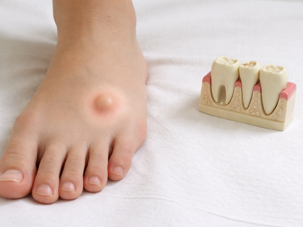
Can Teeth Grow on Your Feet? Causes and What to Do
No, teeth can’t grow on feet. Learn causes of tooth-like bumps, red flags, and next steps for diagnosis.
Do Milk Teeth Grow Back? Timelines, What to Do, Red Flags
Learn if milk teeth regrow, typical timelines for permanent teeth, what to do after loss or injury, and red flags.
Can Milk Teeth Grow Twice? Replacement vs True Regrowth
Can milk teeth grow twice? Learn why baby teeth usually only get replaced, not regrown, and what to do if one falls out