No, teeth cannot grow on your feet. Not even close. True teeth require a very specific set of tooth-forming tissues (dental lamina, ameloblasts, odontoblasts) that are only present in the oral region during embryonic development. The skin, bone, and soft tissue of your feet simply do not have the biological machinery to produce enamel or dentin, ever. If something hard, lumpy, or unusual is showing up on your foot and it looks vaguely tooth-like, it is something else entirely, and figuring out what it actually is matters.
Can Teeth Grow on Your Feet? Causes and What to Do

Marcus Holloway
6 Jun 2026
Why teeth literally cannot form on your feet
Teeth are not just calcium deposits that happen anywhere the body feels like making them. They form through a tightly choreographed process during embryonic development involving two very specific tissue types: oral ectoderm (which becomes the dental lamina and eventually produces enamel via specialized cells called ameloblasts) and neural-crest-derived mesenchyme (which produces dentin via cells called odontoblasts). These two tissue types have to signal back and forth to each other through specific molecular pathways for a tooth to develop at all.
Your feet have neither of these tissue types in the right configuration. The skin on your sole is just skin. The bones in your foot are not odontogenic (tooth-forming) structures. There is no dental lamina hiding in your arch waiting to activate. The epithelial-mesenchymal signaling program required for tooth organogenesis is essentially exclusive to the craniofacial region during a very narrow developmental window. After that window closes, even your mouth loses the capacity to spontaneously generate new teeth.
For context, the rare documented cases of ectopic (out-of-place) teeth in humans involve locations like the maxillary sinus, nasal cavity, orbit, palate, mandibular condyle, and chin. These are all craniofacial and bony sites that are at least adjacent to the original odontogenic tissue fields. Even in those cases, the mechanism is a developmental misdirection of odontogenic tissue during embryogenesis, not spontaneous tooth formation from generic tissue. There are zero credible documented cases of true enamel-and-dentin teeth forming on a foot, hand, or other distant extremity in humans.
So what is that thing on your foot?
This is the genuinely useful part. People describe foot growths as "tooth-like" for a few reasons: they are hard, they protrude, they are white or pale, or they appeared seemingly out of nowhere. Here are the actual conditions that fit that description:
Calluses and corns
These are thickened accumulations of keratin (dead skin protein) caused by repeated pressure or friction. A corn, especially a hard corn on a toe, can feel almost like a dense, pointed nodule and can look pale or yellowish. After paring the surface, a callus will reveal smooth translucent skin underneath, while a wart will show tiny black or red dots (thrombosed capillaries). These are the most common explanation for hard, raised lumps on feet and are not dangerous, though they can be painful.
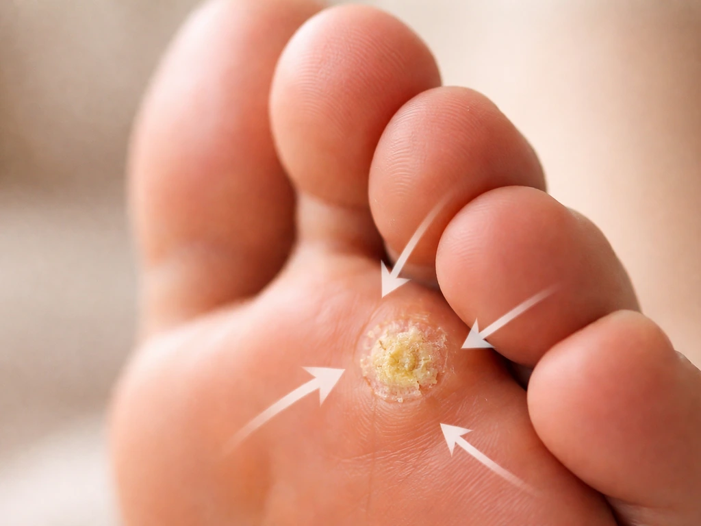
Plantar warts
Caused by human papillomavirus (HPV), plantar warts grow on the sole of the foot and can become quite hard and thick due to pressure from walking. On dermoscopy, they show characteristic red or black dots from tiny blood vessels, which is the fastest way to tell them apart from calluses. They are not teeth, but their hard, lumpy, sometimes clustered appearance is enough to confuse people.
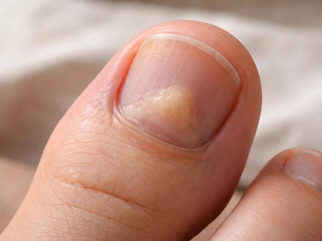
Subungual exostosis
This one genuinely looks like something is growing out of or under your toe. A subungual exostosis is a bony outgrowth from the distal phalanx (the end of the toe bone), usually under the toenail. It can produce a hard, enlarging nodule that pushes the nail up, causes pain, and can even ulcerate the overlying skin. If you have a hard lump near your toenail that is getting bigger and hurts, this is high on the list. An X-ray will confirm the bony origin.
Calcinosis cutis
This is the condition most likely to be described as truly "tooth-like" because it involves actual calcium deposits forming in the skin or subcutaneous tissue. Lesions feel rock-hard, can appear white or chalky when they break through the skin surface, and can occur on the feet and extremities. A published case report describes a solitary hard nodule on the foot present since birth due to calcinosis cutis, which required imaging and pathology to diagnose. The calcium here is not organized into enamel or dentin and has nothing to do with tooth tissue, but it is easy to see why someone would find it alarming.
Cysts, foreign body reactions, and other masses
Epidermoid cysts, inclusion cysts (especially after an old wound or puncture), and foreign body granulomas can all produce firm nodules under the foot skin. In web spaces of toes, pilonidal-type processes involving hair and foreign body deposition can create sinus tracts and lumps that seem to appear from nowhere. None of these are teeth, but they do need evaluation.
Red flags: when to stop waiting and see someone
Most unusual foot growths are benign, but some of these red flags mean you need professional evaluation promptly, not eventually:
- Rapid growth over days to weeks
- The lesion is not healing or is getting larger despite not being touched
- Bleeding from the growth, especially without injury
- Drainage of fluid, pus, or chalky material from the skin
- Spreading redness, warmth, or swelling around the area (possible infection)
- Fever or chills alongside a painful foot lump (urgent: could indicate deep infection or osteomyelitis)
- Numbness, tingling, or loss of sensation in the foot near the growth
- Ulceration of the overlying skin
- A hard lump under or near the toenail that is lifting the nail or causing escalating pain
If osteomyelitis (bone infection) is even a remote possibility, such as when you have swelling, warmth, tenderness over the bone, and systemic symptoms like fever, that requires urgent evaluation. Delays can lead to serious joint and bone damage. See a doctor or go to urgent care the same day.
Who to see and what they will do
A podiatrist is your best first call for most foot growths, especially bony or subungual ones. A dermatologist is appropriate for skin-based lesions. For anything suspicious for malignancy, your primary care doctor can coordinate the right referral. In practice, here is what the clinical workup usually looks like:
- Visual exam and history: when did it appear, has it changed, any pain, trauma, or systemic illness?
- Paring the lesion: for corns and warts, gentle paring reveals important diagnostic clues without any equipment
- Dermoscopy: a handheld magnifier used by dermatologists to see vascular patterns, especially useful for warts vs. calluses
- X-ray: ordered when a bony origin is suspected (subungual exostosis, osteomyelitis, calcinosis with deeper involvement)
- MRI or CT: for deeper soft tissue or bone involvement when X-ray is not enough
- Biopsy: when malignancy cannot be excluded, or when the lesion type is uncertain, a punch or excisional biopsy sends tissue to pathology for a definitive answer
Can teeth regrow anywhere in humans? The honest limits
Since this question often comes from a broader curiosity about tooth regeneration, here is the straight answer: in humans, enamel cannot regenerate at all after it forms. Once ameloblasts complete their job during tooth development, they die. The enamel they created is acellular, meaning there are no living cells left in it to rebuild it. Dentin has slightly more potential because dental pulp stem cells (DPSCs) can produce odontoblast-like cells after injury, but this is a limited repair response, not true regrowth. Cementum (which covers tooth roots) has some limited capacity to reform after disease-induced resorption.
The adult human mouth cannot grow a third set of teeth. After the dental lamina disintegrates following the eruption of permanent teeth, the structural blueprint for tooth regeneration is essentially gone. Researchers are actively working on ways to reactivate dental stem cells for tooth regeneration, but as of today, this remains experimental. The idea that teeth could regenerate anywhere other than the mouth, let alone on your feet, has no biological basis whatsoever. Teeth also cannot grow under your tongue, because true tooth formation requires the same specialized embryonic oral tissues and signaling that are not present there can teeth grow under your tongue.
What animals can do that humans cannot
It is worth addressing the animal angle, because the internet sometimes mixes up human dental biology with what happens in other species. Sharks are the most famous example: they continuously regenerate teeth throughout their lives because they retain an active dental lamina with Sox2-positive stem cell populations that keep producing new tooth generations. Reptiles like snakes and lizards are polyphyodonts (multiple tooth sets) for the same reason: their dental lamina remains intact and functional.
Humans, along with most other mammals, are diphyodonts: two sets of teeth (baby and adult), and then the dental lamina disintegrates. This is why even in animals with impressive tooth regeneration abilities, that regeneration happens exclusively in the oral cavity through an active dental lamina system. Not on their fins. Not on their tails. Not on their feet. The regenerative biology is site-specific, oral-tissue-specific, and entirely absent in foot tissue across virtually all known vertebrates.
This is also relevant if you have seen claims about teratomas (tumors that can contain hair, teeth, and other tissue types from multiple germ layers). Teratomas can technically contain tooth-like structures, but they are rare developmental tumors, not teeth "growing" anywhere normal, and they require medical diagnosis and management. They are not something to self-diagnose from a foot lump.
What to actually do today

If something unusual is on your foot right now, here is a practical action plan:
- Take a clear photo of the lesion in good lighting today. Track it by measuring or photographing it weekly so you can tell a clinician whether it has changed.
- Do not try to cut, dig out, or shave down a hard growth at home, especially anything near a toenail or under the skin. You risk infection.
- Do not put off evaluation if you have any of the red flags listed above. Same-day care is appropriate for fever, spreading redness, or signs of deep infection.
- For a non-urgent but persistent or growing lesion, book with a podiatrist or dermatologist within the next week or two.
- Before your appointment, write down: when you first noticed it, whether it has changed in size or appearance, any pain or discharge, any recent foot trauma or injury, and any relevant health conditions (diabetes, autoimmune conditions, circulatory problems).
- At the appointment, ask directly: what is this, what do I need to rule out, and do I need imaging or a biopsy?
The bottom line is simple: what you are looking at is not a tooth, because teeth cannot form on feet. But that does not mean you should ignore it. Hard, growing, or painful lumps on the foot have real causes that a clinician can identify and treat, often straightforwardly once the right diagnosis is made. Dermatologists often blank" rel="noopener noreferrer">perform skin biopsies when a growth needs diagnosis, and biopsy results depend on appropriate sampling and pathology evaluation. Get it looked at, describe what you see accurately, and let the workup tell you what it actually is.
FAQ
If I can feel something hard on my foot, how can I tell if it is a tooth versus something else?
Not in the way people mean it. If a “tooth-like” bit is actually coming from under the skin or near a nail, it is more often a wart, callus, bony outgrowth, or calcium-containing skin lesion. The only safe way to confirm is an in-person exam, and if it is near bone or a toenail, clinicians commonly use imaging such as an X-ray.
Can I safely cut or pull off a “tooth-like” lump on my foot at home?
Do not try to remove it yourself with cutting, drilling, or burning. For calluses you may damage healthy skin, for warts you can spread the virus, and for bony lesions you can cause bleeding, infection, or worsen pain. If it is thick and painful, it is reasonable to cover it and schedule a podiatry or dermatology visit instead of DIY removal.
What symptoms mean I should see a doctor sooner rather than later for a foot lump?
A good rule is that any foot growth that is rapidly enlarging, ulcerating, draining, changing color quickly, or associated with persistent night pain deserves prompt evaluation. Another practical trigger is functional impact, if it is affecting walking or wearing shoes, since pressure changes can make benign lesions worse and can delay accurate diagnosis.
What information should I collect before my appointment for a hard foot growth?
Take clear photos in good lighting from a couple angles, note the approximate start date, and track changes weekly (size, color, pain, and whether it bleeds). If it is near a toenail, note whether the nail is lifting or deforming. This helps a clinician distinguish skin-based lesions from bony or nail-origin problems quickly.
Which specialist should I see first, podiatry or dermatology, for a “tooth-like” spot?
If the lump is mainly in or under the toenail, a podiatrist is usually the best first call, because subungual exostosis and other bony causes are common there and may need an X-ray. If it is on the sole surface and looks like a rough or clustered growth, dermatology or podiatry can evaluate for warts. If you have diabetes, poor circulation, or immune suppression, earlier assessment is especially important.
If teeth cannot grow on feet, can any treatments make the problem go away without a diagnosis?
Enamel and dentin do not regenerate in humans, so brushing, supplements, or “tooth whitening” products cannot change a foot lump into teeth. However, basic foot care can reduce irritation if the cause is pressure related, like proper shoe fit and offloading, while you get it diagnosed. If it is a wart, the treatment plan is different, so avoid guessing based on appearance alone.
Why do some foot lesions look chalky or “calcium-like” if they are not teeth?
Yes, some non-tooth conditions can mimic teeth because they are hard, white or chalky, and can break through skin. Calcinosis cutis can feel rock-hard, warts can thicken under pressure, and bony lesions under a toenail can produce a firm enlarging bump. Imaging and sometimes pathology are the typical next steps when something is very hard and persistent.
What tests are commonly used to diagnose hard lumps that seem tooth-like on the foot?
If a lesion looks like a bony outgrowth, especially near the nail, an X-ray is often the fastest way to determine whether bone is involved. For skin lesions that are very firm or unusual, clinicians may use ultrasound, blood work if calcium issues are suspected, or biopsy/pathology for calcified or persistent nodules. The workup depends on exam location and how fast it is changing.
Next Articles
Do Milk Teeth Grow Back? Timelines, What to Do, Red Flags
Learn if milk teeth regrow, typical timelines for permanent teeth, what to do after loss or injury, and red flags.
Can Milk Teeth Grow Twice? Replacement vs True Regrowth
Can milk teeth grow twice? Learn why baby teeth usually only get replaced, not regrown, and what to do if one falls out

Why Do Teeth Only Grow Twice? Baby vs Permanent Explained
Why humans have only baby and permanent teeth: eruption timeline, limits of enamel regrowth, and common exceptions to kn