If you've spotted what looks like a tooth growing in a strange spot, whether it's a lump on the roof of the mouth, something poking through in the back of the jaw where no tooth should be, or a weird hard bump in soft tissue, it almost certainly isn't a tooth "regrowing" from scratch. What you're most likely seeing is an existing tooth bud that ended up in the wrong place, an extra tooth that shouldn't be there at all, or a tooth-like growth made of the same cellular material teeth are built from. Human teeth do not regenerate. But they absolutely can erupt in some genuinely strange locations, and that's worth understanding.
Weird Places Teeth Can Grow: Causes, Signs, and Next Steps

Marcus Holloway
20 May 2026
What "teeth in weird places" actually means
The phrase covers a few different things that get lumped together, and it matters which one you're dealing with.
The most common situations are ectopic eruption (a normal tooth that erupts in an abnormal position), supernumerary teeth (extra teeth that shouldn't exist at all), impacted teeth (teeth that stay buried in bone or tissue well past when they should have appeared), retained primary teeth (baby teeth still sitting there when they should have fallen out long ago), and odontomas (benign tooth-like growths made of dental tissue that aren't quite "teeth" but look like them on an X-ray).
Each has its own cause, its own set of symptoms, and its own management path. The one thing they all share is that they're not spontaneous regrowth. They all trace back to existing tooth-forming tissue that went somewhere it wasn't supposed to go.
The most common unusual locations where teeth turn up
Most ectopic or supernumerary teeth show up in predictable "weird" spots, and the location usually tells you something about the likely cause.
The palate (roof of the mouth)
This is probably the most common unusual eruption site people notice at home. Palatally displaced canines are seen in roughly 1 to 2 percent of the general population and are even more common in adolescent orthodontic patients, around 3 to 5 percent. Instead of dropping down into the arch like a normal canine, the tooth bud migrates toward the roof of the mouth and either stays buried or erupts through palatal tissue. You might feel a hard ridge on the palate behind the front teeth, or notice that a canine in the upper arch never came in at all.
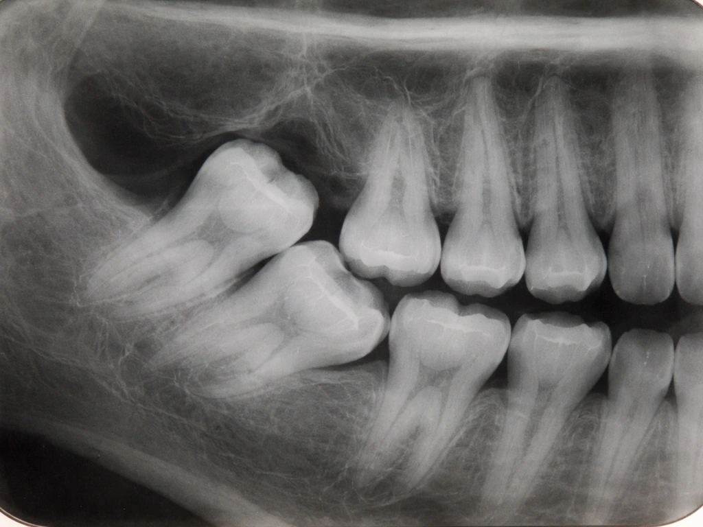
Buried within the jaw (impacted teeth)
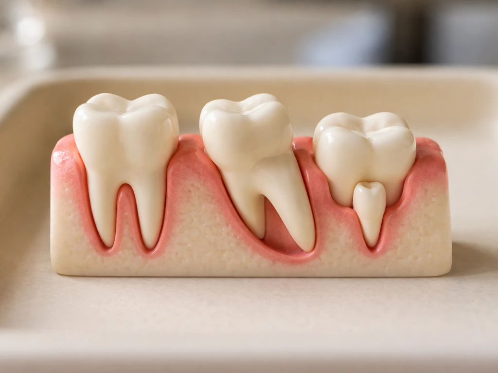
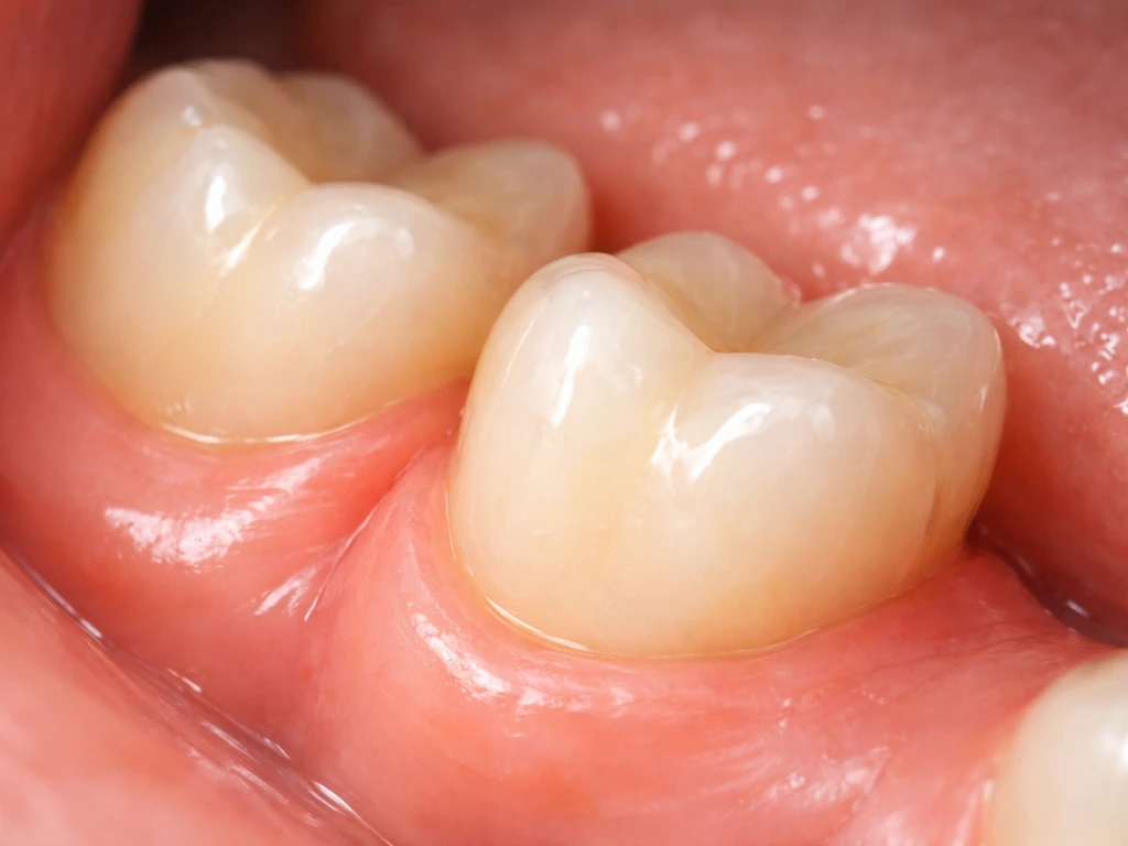
An impacted tooth is one that remains fully or partially embedded in the jawbone or mucosa for more than two years past its normal eruption window. Wisdom teeth are the most familiar example, but canines, premolars, and even molars can all become impacted. The tooth is there on an X-ray, but it's either stuck against another tooth, angled in the wrong direction, or simply doesn't have enough space to break through.
The nasal cavity and nasal septum
This one surprises people, but it's documented. Nasal teeth, or ectopic teeth that erupt into the nasal cavity, are rare but real. Published case reports describe teeth appearing in the nasal floor, the nasal septum, and even further into the nasal passage. They can look like a yellowish-white mass surrounded by granulation tissue and debris on exam. Imaging, typically a CT scan, shows the tooth-like structure clearly. The fact that a tooth can wind up here comes down to the anatomy: the upper jaw's tooth-forming tissue sits very close to the floor of the nasal cavity during development, especially near the front teeth.
The maxillary sinus
Ectopic teeth have been found projecting into the maxillary sinus, the air-filled cavity just above the upper back teeth. Cases have presented with chronic sinusitis, recurrent nosebleeds, headaches, facial numbness, and in at least one published report, recurrent haemoptysis (coughing up blood). On a CT scan these can be tricky to interpret because benign odontogenic tumors like odontomas and calcifying cysts can look similar, which is exactly why imaging with proper diagnosis matters and not just "it looks like a tooth, let's pull it."
Soft tissue and unusual jawbone positions
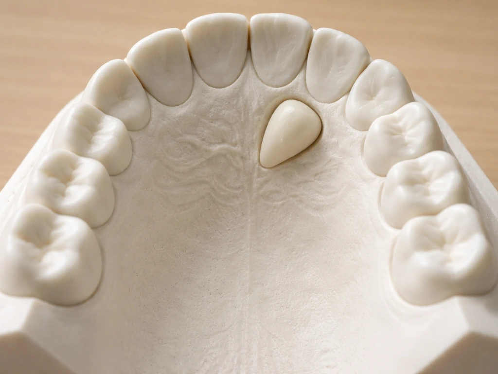
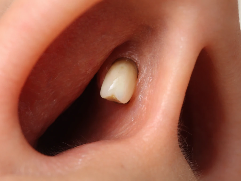
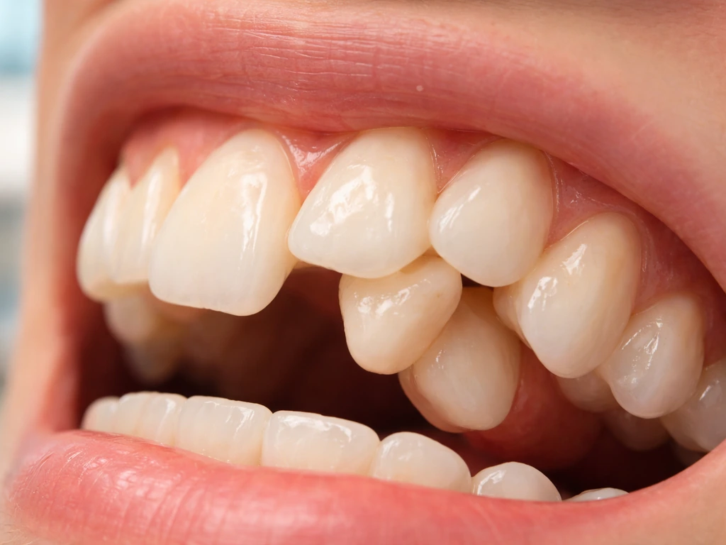
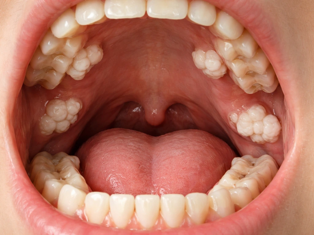
Supernumerary teeth can erupt through gingival tissue in spots where no tooth is expected, such as behind the last molar, between the front teeth (a midline extra tooth called a mesiodens is a classic example), or even through tissue on the lingual side of the arch. Odontomas, which are hamartoma-type lesions made of enamel, dentin, cementum, and pulp, are usually found in the jawbone on routine X-rays rather than spotted visually. They're not tumors in the conventional sense but they can block normal teeth from erupting.
Why it happens: the biology behind teeth ending up in the wrong place
There are four main explanations, and in many cases more than one factor is at play.
Developmental disturbances during tooth formation
Teeth form from a strip of specialized tissue called the dental lamina. If that lamina becomes overactive, it can produce extra tooth buds, which is what leads to supernumerary teeth and hyperdontia. If it produces a bud in an abnormal position or orientation, ectopic eruption is the result. Cleft lip and palate is a well-documented developmental condition associated with both ectopic eruption and supernumerary teeth, partly because the structural disruption to the palate changes where tooth-forming tissue sits during early development.
Genetics and syndromes
Supernumerary teeth have a clear genetic dimension. They appear at higher rates in families with cleidocranial dysplasia, Gardner's syndrome (familial adenomatous polyposis), and a handful of other conditions. If you have hyperdontia with no known syndrome, hereditary factors and dental lamina hyperactivity are the most likely contributors. Worth noting: many websites throw around a long list of syndromes associated with extra teeth, but the research is actually more selective. Only about eight genetic syndromes have strong published evidence for the association.
Trauma and displacement
A direct blow to the face during childhood can physically displace a tooth bud before it has erupted. One published BMJ Case Reports case documented an intranasal tooth that erupted a full year after maxillofacial trauma in a child, with CT imaging showing the bud had been pushed into the nasal floor at the time of injury.
A 2014 BMJ Case Reports report described an intranasal ectopic tooth that erupted one year after maxillofacial trauma in a child, after the tooth bud was displaced to the nasal floor intranasal ectopic tooth erupted a full year after maxillofacial trauma. This is a particularly important cause to understand for parents: if a child has had a significant facial injury, ectopic eruption can appear later and the connection to the original trauma isn't always obvious.
Crowding, obstruction, and path-of-eruption problems
Sometimes a tooth erupts in the wrong place simply because its normal path was blocked. Dense bone, persistent retained primary teeth, cysts, or crowding can all redirect a tooth bud. In ectopic eruption specifically, the abnormal path can cause the ectopic tooth to resorb (eat away at) the root of a neighboring primary tooth, leading to premature loss of that baby tooth and malposition of the permanent one.
Regrowth vs eruption: what teeth can and can't actually do
This is the myth-busting part. When someone says "a tooth grew back" or "a tooth is growing somewhere new," they almost always mean eruption, not regeneration. These are very different things. Eruption means an existing tooth (or tooth-like structure) that was already formed is moving through tissue and becoming visible.
Regeneration would mean new dental tissue forming from nothing after the original is gone or damaged. Human teeth cannot regenerate. Human tooth growth happens at dental sites, and teeth generally do not grow on fingers. Enamel, once formed and lost, cannot be replaced by the body.
Dentin and pulp have very limited repair capacity under the right conditions, but nothing close to regrowth. A whole new tooth cannot spontaneously form after permanent teeth are established.
Some animals can do this. Fish and many reptiles grow new teeth throughout their lives. Mice have incisors that continuously grow and renew, which makes them a useful model for dental stem cell research. But mammals, including humans, largely lost that capacity through evolution. Current dental regeneration research is focused on stem cell therapies and tissue engineering in lab and early clinical settings, not anything naturally occurring in a living human mouth. So if you're reading that "teeth can regrow anywhere in the body," that's not a real description of human biology.
What people sometimes confuse for regrowth: a wisdom tooth erupting in adulthood (it was always there, just buried), a retained baby tooth finally falling out and revealing a permanent tooth underneath that's been there for years, an odontoma becoming visible as it enlarges, or a supernumerary tooth erupting at an unexpected time. All of these involve pre-existing dental tissue moving or becoming visible. None of them are regeneration.
Symptoms and red flags: when to take it seriously
Some ectopic or supernumerary teeth are completely asymptomatic and are found incidentally on a routine X-ray. Others cause real problems. The symptoms depend heavily on where the tooth is and what it's pressing against.
For teeth in or near the nasal cavity or sinus, symptoms can include nasal obstruction, foul-smelling discharge from one nostril, recurrent nosebleeds, facial pain or pressure, headaches, and in some cases nasolacrimal duct obstruction (which causes excessive tearing or epiphora). These symptoms are easy to mistake for a sinus infection or allergies, and if they're chronic or one-sided, it's worth mentioning to a doctor that an ectopic tooth is a possibility, even if it sounds far-fetched.
For teeth in the jaw or palate, common signs are a permanent tooth that simply never came in (non-eruption of a permanent tooth is the most frequently reported complaint in CBCT studies of supernumerary teeth), crowding or shifting of adjacent teeth, a hard lump on the palate or gum, or discomfort and pressure in a specific area.
The following symptoms are urgent and mean you should contact a dental or medical provider the same day, not wait for a scheduled appointment:
- Swelling in the face, jaw, or neck
- Pus or drainage from the gum or a lump
- Fever alongside jaw or oral pain
- Worsening pain that doesn't respond to over-the-counter pain relief
- Difficulty opening your mouth, swallowing, or breathing
- Sudden changes in vision or significantly worsening eye symptoms (if there's a known or suspected ectopic tooth near the orbit or nasolacrimal duct)
An infection around an impacted or ectopic tooth can spread rapidly. These aren't "wait and see" situations.
How dentists figure out what's going on
Diagnosis starts with a clinical exam: the dentist looks at what's visible, palpates the tissues, notes what teeth are and aren't present in the arch, and asks about symptoms and history including any prior facial trauma. If the suspected issue is in the jaw, a panoramic X-ray is typically the first imaging step. It gives a broad view of all teeth, their positions, root development, and anything unusual in the jawbone.
If the panoramic image shows something that needs more precise localization, or if the clinical picture is complex, the next step is usually cone beam CT (CBCT). CBCT gives a three-dimensional view of the tooth and its relationship to surrounding structures, including nerves, adjacent roots, the nasal floor, and the sinus. For surgical planning of impacted or ectopic teeth, this detail is often essential. CBCT isn't ordered for every case, though. The clinical standard is to justify it based on what was found on initial exam and conventional imaging, not as a first step.
When a tooth-like finding is in or near the nasal cavity or sinus, the differential diagnosis gets broader. A calcified mass in the nasal passage could be a rhinolith (mineralized foreign body), an odontoma, a calcifying odontogenic cyst, a benign or malignant tumor, or an inflammatory lesion from conditions like fungal infection or granulomatous disease. Imaging characteristics on CT help distinguish these, but in some cases a tissue biopsy or surgical removal with pathology is the only way to be sure. This is part of why an ENT specialist often gets involved when the finding is in the nasal cavity or sinus.
Treatment options: from watchful waiting to surgical removal
Management depends on what type of abnormal tooth situation you're dealing with, where it is, and whether it's causing or risking damage to adjacent structures.
| Situation | Typical management approach | Specialist involved |
|---|---|---|
| Asymptomatic impacted tooth (e.g., wisdom tooth) with adequate space | Monitoring with periodic X-rays; extraction if problems develop | General dentist or oral surgeon |
| Impacted canine or premolar in a younger patient | Orthodontic space creation + surgical exposure + guided eruption | Orthodontist and oral surgeon |
| Supernumerary tooth blocking normal eruption | Extraction of supernumerary tooth; orthodontic follow-up to allow normal eruption | Oral surgeon and orthodontist |
| Odontoma in the jawbone | Surgical removal, especially if blocking eruption; monitoring if very small and asymptomatic | Oral surgeon; pathology review of specimen |
| Ectopic tooth in nasal cavity or sinus | Surgical removal (often endoscopic for nasal teeth); ENT involvement for nasal/sinus location | ENT surgeon, oral and maxillofacial surgeon |
| Palatally displaced canine | Orthodontic treatment to open space; surgical exposure; bonded attachment and traction | Orthodontist and oral surgeon |
The general principle is: if an ectopic or supernumerary tooth is causing symptoms, damaging adjacent teeth, blocking normal eruption, or poses infection risk, it should be addressed. If it's completely asymptomatic, small, not growing, and not near anything critical, monitoring is reasonable. Early detection consistently leads to better outcomes, particularly in children, because there's more flexibility in the bone and more time to guide eruption before the arch is fully developed.
For parents: if your child's permanent tooth hasn't come in within about six months of the baby tooth falling out, that's worth bringing up at a dental appointment. Delayed eruption is the most common presenting complaint in patients who turn out to have a supernumerary tooth blocking the path.
For adults: a tooth growing somewhere unexpected is not a dental emergency in most cases, but it does need evaluation. Don't wait years hoping it resolves on its own. A panoramic X-ray gives your dentist a fast, low-cost first look at what's happening, and from there you'll know whether it needs specialist referral or just monitoring.
What to do right now if you're worried
- If there's pain, swelling, fever, drainage, or difficulty swallowing, contact a dentist or emergency dental clinic today. These symptoms suggest infection, which moves fast.
- If you've found a hard lump, a visible extra tooth, or a tooth that appeared somewhere it shouldn't be but you have no acute symptoms, call your regular dentist and get a clinical exam and panoramic X-ray scheduled within a week or two.
- If a child's expected permanent tooth hasn't erupted six months after the baby tooth came out, bring it up at the next dental visit and ask about imaging.
- If you have chronic one-sided nasal symptoms, mention the possibility of a nasal or sinus tooth to your physician or dentist, especially if you have a history of facial trauma or a known supernumerary tooth.
- Bring up any relevant history: past facial injuries, family history of extra teeth, or any known syndromes. This context helps narrow the diagnosis quickly.
- If your dentist refers you to an oral surgeon or orthodontist, follow through. These cases often need more than one specialist, and delays can mean more complicated treatment later.
The broader picture here is that "teeth growing in weird places" is a real phenomenon, not internet folklore, but it's almost never spontaneous regrowth. A condition often discussed online as a “disease where teeth grow everywhere” is still best understood as ectopic eruption or extra teeth rather than true regeneration teeth growing in weird places. It's existing dental tissue that ended up somewhere abnormal during development, after trauma, or because its normal path was blocked. Understanding the difference between eruption and regeneration helps cut through a lot of the confusion online.
Related curiosities, like whether teeth can appear elsewhere in the body in a more extreme sense, or what it means when teeth seem to grow on top of existing ones, are worth exploring separately but follow the same underlying biology. Some people also ask whether a tooth can grow under another tooth, which usually ends up being a case of eruption or an impacted or supernumerary tooth rather than true regrowth.
That includes conditions like hyperdontia, a disease where teeth grow everywhere. In a more extreme sense, this is why people ask whether teeth can grow in other parts of the body. The practical bottom line: get it examined, get imaging, and don't wait if you have any of the urgent symptoms above.
FAQ
How can I tell if what I see is an erupted tooth versus a non-tooth lump like a cyst or tumor?
Location and feel help, but the reliable way is imaging. Teeth-related findings usually show a tooth-like calcified structure on X-ray or CT and often have an associated missing or malpositioned neighboring tooth. A soft, fluctuant bump, rapidly enlarging tissue, or one that bleeds easily needs prompt evaluation because those patterns can fit cysts or tumors, not erupted dental tissue.
If the spot looks like a tooth in the palate or gum, should I expect pain or infection right away?
Not necessarily. Many ectopic or supernumerary teeth are found incidentally and cause no symptoms early. However, if the area is tender, there is swelling, bad taste or drainage, or fever develops, that suggests secondary infection around the abnormal structure and warrants same-day medical or dental care.
Could a tooth growing near the nasal area be mistaken for allergies or a sinus infection?
Yes, it commonly is. A one-sided pattern, recurrent or foul-smelling discharge, repeated nosebleeds, or persistent facial pressure that does not match typical allergy triggers should raise suspicion for a local structural cause. If symptoms are chronic or primarily unilateral, tell the clinician that an ectopic dental origin has been considered so appropriate imaging can be ordered.
What is the safest first imaging test for adults with a tooth in an unexpected location?
A panoramic X-ray is often the first step because it provides a broad survey of teeth and jaw structures at relatively low cost and radiation compared with 3D scans. If the finding involves proximity to the sinus, nasal floor, or nerves, or if the panoramic image is unclear, cone beam CT is typically used next for precise localization.
Do impacted teeth or supernumerary teeth always need removal?
No. If they are small, not growing, not blocking eruption, and not threatening adjacent roots, nerves, or other critical structures, monitoring may be reasonable. Treatment becomes more likely if there is pain, infection risk, damage to neighboring teeth, impaction-related symptoms, or obstruction of normal eruption.
In children, when should delayed eruption trigger concern for a supernumerary tooth?
A practical rule is to discuss it with a dentist if the permanent tooth has not appeared within about six months after the baby tooth falls out. Delayed eruption is a common reason supernumerary teeth are discovered, and earlier evaluation gives more options for guiding the tooth into the arch.
If I had facial trauma as a child, could an ectopic tooth appear years later?
It can. Trauma can displace a tooth bud before eruption, and the abnormal position may only become apparent when that tooth would normally erupt. If you have a history of significant maxillofacial injury and a new tooth-like structure appears in an unexpected site, mention the trauma timeline, because it can change the diagnostic reasoning.
Could a retained baby tooth be causing an ectopic eruption or tooth misalignment?
Yes. A retained primary tooth can block the normal eruption path of the permanent successor, leading to delayed eruption, abnormal angulation, or ectopic eruption. In some cases, the developing permanent tooth can also resorb the root of the retained baby tooth, so the status of both teeth matters, not just the new-looking bump.
What are the urgent red flags that should not wait for a routine appointment?
Seek same-day care if there are signs of spreading infection (rapid swelling, fever, worsening pain), trouble breathing or swallowing, or significant facial swelling with systemic symptoms. For lesions near the sinus or nasal cavity, persistent high-grade symptoms like significant one-sided nosebleeds or severe worsening facial pain also deserve urgent evaluation.
Can I safely try to remove or poke the tooth-like bump at home?
No. Even if it appears firm, it may be an ectopic tooth or a tooth-like calcified mass embedded in tissue. Attempting to remove it can cause infection, bleeding, and delayed diagnosis. The correct next step is assessment with an exam and appropriate imaging.
If imaging shows something tooth-shaped in the sinus or nasal passage, why might a biopsy be needed?
Because several non-dental conditions can look similar on scans, including certain cysts, tumors, fungal or inflammatory lesions, and calcified foreign body problems. When imaging characteristics do not confidently narrow it to a dental origin, pathology from surgical removal or biopsy may be the only way to confirm the diagnosis and guide treatment.
What does it mean when symptoms are worse on one side of the face or nose?
One-sided, localized symptoms are more suggestive of a structural or localized lesion than generalized causes like typical viral illness or common allergies. Unilateral chronic pressure, recurrent discharge, or repeated bleeding should prompt evaluation for a specific local source, including ectopic dental tissue.
Next Articles
Can a Tooth Grow Under Another Tooth? What It Means
Yes, teeth can erupt behind or under others due to impaction or extra teeth, but true regrowth is rare. What to do.
Teeth That Grow on Top of Teeth: Causes and What to Do
Causes of teeth that appear over others, true regrowth limits, symptoms, diagnosis steps, and treatment options by age.
Disease Where Teeth Grow Everywhere: Causes, Diagnosis, Treatment
Learn why teeth-like growth can appear everywhere, how it’s diagnosed with imaging, and what treatments are possible vs