Yes, gum tissue can grow over a bone graft site, but it does not always do so on its own, and it does not happen the same way for every patient or every type of graft. When the healing goes as planned, soft tissue slowly migrates and closes over the grafted area within a few weeks. When it does not close properly, you get what clinicians call a "dehiscence" or exposure, and that needs active management rather than a wait-and-see approach. So the short biological reality is: gum regrowth over a bone graft is possible and common, but it is not guaranteed, and exposed bone after a graft is a real complication that your oral surgeon needs to know about promptly.
Does Gum Grow Over Bone Graft? What to Expect and Next Steps

Marcus Holloway
2 Jun 2026

What "exposed bone" actually means after a graft
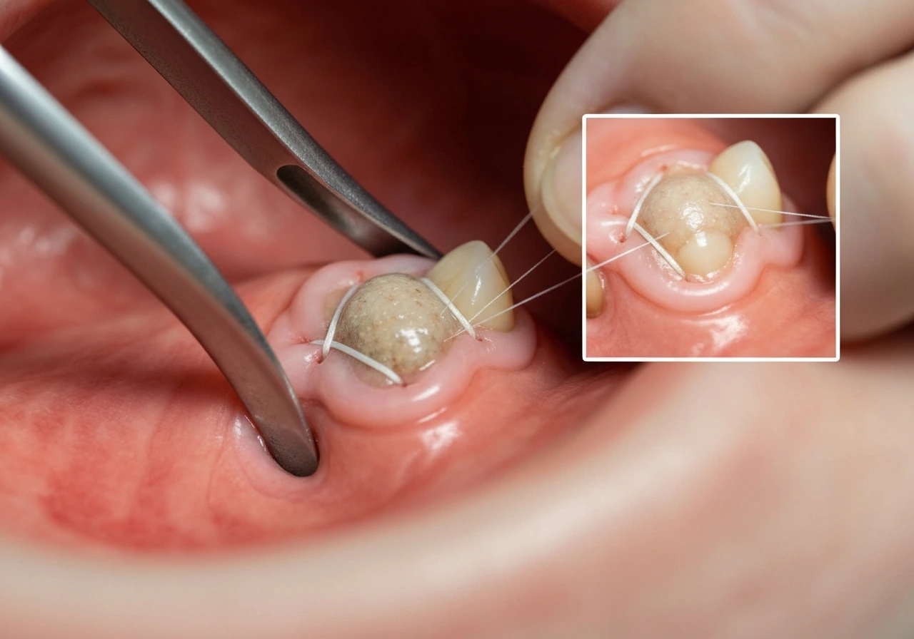
After a bone graft, your surgeon places graft material (and often a barrier membrane on top of it) into the defect, then closes the gum tissue over everything with sutures. The goal is a watertight, primary closure where soft tissue seals the whole site within the first two weeks. When that seal breaks down, you can end up with visible graft particles, membrane material, or even raw bone peeking through the gum. This is called a dehiscence, and it happens more often than most patients realize. Studies on guided bone regeneration (GBR) procedures put the rate of membrane exposure alone at roughly 22 to 23 percent, meaning nearly one in four GBR sites experiences some form of soft-tissue breakdown. So if you are looking at something white or chalky through your gum tissue right now, you are not alone and you did not necessarily do anything wrong.
The most common reasons this happens include tension in the flap at the time of surgery (the gum was stretched too far to cover the graft volume), swelling in the days after surgery that pulls sutures apart, trauma to the site from eating or brushing too aggressively, infection, or the graft volume being too large to allow tension-free closure. The type of membrane matters too. Non-resorbable membranes, like dense PTFE (dPTFE), are more likely to stay exposed because they do not dissolve on their own, while resorbable collagen membranes can break down and actually act as a temporary cover even after the gum pulls back slightly.
Week-by-week: what healing actually looks like
Healing after a bone graft is not linear, and the gum tissue closing over the site does not look like a zipper. Here is a practical sense of what to expect at each stage.
| Timeframe | What you may see | What is happening biologically |
|---|---|---|
| Days 1 to 3 | Swelling, bruising, mild bleeding, sutures intact | Blood clot forming; soft-tissue flap holding closure; inflammation peaks |
| Days 4 to 7 | Swelling starts to ease; sutures may loosen; possible white film near wound edge | Clot matures; early granulation tissue begins forming under the flap |
| Week 2 | Sutures dissolving or removed; wound edges should be pink and approximated | Soft-tissue seal is most critical here; the first 15 days are the key stabilization window |
| Weeks 3 to 4 | Gum tissue looks more normal; any minor gap may start to fill in | Epithelial migration and connective tissue remodeling; graft material stabilizing |
| Weeks 6 to 12 | Site appears fully covered in most uncomplicated cases; some firmness or color difference normal | Bone formation underway beneath the soft tissue; membrane either resorbing or scheduled for removal |
| Months 4 to 6 | Re-entry or implant placement evaluation; site assessed for bone volume gained | Bone graft maturation; for non-resorbable membranes, this is when they are typically removed |
The first two weeks are the most important window. Research on GBR specifically flags the first 15 days after a procedure as critical for maintaining the soft-tissue seal. If the gum stays closed during that window, the rest of the healing process is far more predictable. If you notice the site opening up in week one or two, that is the time to call, not wait until your next scheduled appointment.
What makes the difference between gum that closes vs. gum that does not
Not every exposed bone situation ends the same way. If you are specifically wondering about whether gums grow to fill gaps after an exposure or dehiscence, the key idea is that closure is possible but not guaranteed and depends on the seal, membrane type, and how early it is managed do gums grow to fill gaps. Several factors influence whether your gum tissue will migrate over the graft on its own, with antiseptic help, or whether it needs surgical correction.
Graft type and membrane choice
Resorbable collagen membranes are more forgiving when exposure occurs. Even if the gum pulls back slightly, the collagen can act as a biological scaffold while tissue slowly migrates over it. Non-resorbable membranes like dPTFE cannot do this. When a dPTFE membrane becomes exposed, it creates a direct bacterial pathway into the graft site, which can compromise bone formation underneath. The clinical approach for resorbable membrane exposure is often aggressive antiseptic management and monitoring. Authoritative ITI Academy module narration on GBR notes that dehiscence can expose an applied membrane, and if that membrane is resorbable the exposed membrane and the soft-tissue dehiscence are managed with mechanical and antiseptic plaque control early stabilization concept of maintaining the soft-tissue seal for about the first 15 days. For non-resorbable membranes, the decision of whether to remove the membrane early is more urgent.
Size and location of the defect
Small dehiscences, a millimeter or two of opening at the wound edge, often resolve with careful antiseptic management. Larger defects, especially those exposing a significant area of graft material, are much less likely to self-close. Posterior sites also tend to be harder to manage because access for home care is limited and the cheek puts pressure on the site when chewing.
Surgical technique and flap design
Primary closure without tension is the gold standard. When a surgeon has to stretch the flap aggressively to cover a large graft volume, the sutures are fighting against tissue tension the moment the local anesthetic wears off. Experienced surgeons use releasing incisions to give the flap more mobility, but even then, very large grafts carry a higher dehiscence risk.
Patient-specific factors
- Smoking: significantly impairs blood flow to healing tissue and dramatically increases dehiscence and graft failure rates
- Uncontrolled diabetes: slows wound healing and increases infection risk at the site
- Thin gum biotype: patients with naturally thin, delicate gum tissue have less tissue volume to work with and are more prone to recession and exposure
- Poor oral hygiene: plaque accumulation at a dehisced site allows bacteria to penetrate and cause infection, which is one of the main reasons small exposures turn into big problems
- Certain medications: blood thinners, immunosuppressants, and bisphosphonates can all affect healing and graft outcomes
Warning signs that need a same-day or urgent call
Some discomfort and odd appearance are normal after a bone graft. These warning signs are not normal and mean you should contact your oral surgeon today, not at your next scheduled visit.
- A foul taste or smell coming from the graft site, which suggests bacterial contamination and possible infection
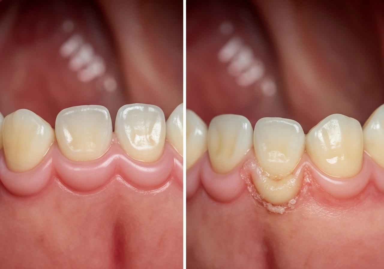
- Visible white, chalky, or granular material that you can see and feel protruding through the gum (exposed graft particles or membrane)
- Pain that is getting worse after day 3 or 4 instead of gradually improving
- Significant swelling, redness, or warmth spreading beyond the immediate graft site
- Pus or discharge from the wound
- Fever above 101 degrees Fahrenheit in the days following surgery
- A suture line that has completely opened and you can see clearly into the wound
- The sensation of something hard and loose moving in the site
A small amount of white tissue at the wound edge is often just normal healing granulation tissue or a dissolving suture, not exposed bone. But if you are unsure, call anyway. Your surgeon would far rather reassure you over the phone than see a complication that was ignored for a week.
What your dental team will likely do next
When you show up with a dehiscence or exposed graft material, the response from your surgeon depends on how bad the exposure is and what type of graft and membrane were used. Here is what the management ladder typically looks like.
For minor dehiscence with resorbable membrane or small graft exposure
The most common approach is a strict antiseptic protocol. That usually means chlorhexidine gel or rinse applied twice daily directly to the area, possibly combined with a course of systemic antibiotics to prevent bacterial invasion. Your surgeon will likely schedule you for weekly check-ins rather than waiting for your standard follow-up. Research on GBR dehiscences consistently shows that most small exposures resolve completely with this type of protocol, with very few cases requiring more extensive intervention.
For non-resorbable membrane exposure
When a dPTFE or other non-resorbable membrane is exposed, the decision your surgeon faces is whether to remove the membrane early and accept potentially reduced bone gain, or to attempt to maintain the site with antiseptics and close monitoring. The longer a non-resorbable membrane stays exposed, the more bacterial contamination risks the underlying bone formation. Some surgeons will attempt re-closure of the flap over the membrane if the exposure is caught early. Others will remove the membrane but leave the graft material in place if the site looks healthy underneath.
For larger or infected exposures
In the most severe cases, where infection is present and the graft material is compromised, your surgeon may need to remove the graft and membrane entirely, debride the area, and allow healing by secondary intention before planning a second grafting procedure. This is not common, but it happens, and it is not the end of the road for your implant or restoration plan. It means a longer timeline, not a failed outcome forever.
Soft-tissue grafting as a follow-up
Once the initial bone grafting heals successfully (even if it took an extra round of management), some patients still end up with thin or insufficient gum tissue over the site. A connective tissue graft or free gingival graft taken from the palate can be placed to build up the soft tissue before or alongside implant placement. This is a separate, less complex procedure than the bone graft itself, and it dramatically improves the long-term health of the tissue around an implant.
How to care for the site right now
What you do at home in the weeks following a bone graft has a real impact on whether that gum tissue stays closed. These are not just standard post-op suggestions. They directly affect whether soft tissue holds over the graft.
- Do not touch the site with your tongue, fingers, or any instrument. Mechanical disturbance of the clot and early tissue seal in week one or two is one of the most common avoidable causes of dehiscence.
- Use chlorhexidine rinse exactly as directed. Most surgeons prescribe a 0.12 percent chlorhexidine gluconate rinse for the first one to two weeks. Do not skip it, and do not rinse aggressively, just gentle swishing.
- Eat soft foods on the opposite side of the mouth. Hard, crunchy, or chewy foods create pressure and movement right at the graft site. Stick with scrambled eggs, yogurt, soup, and similar foods for at least two weeks.
- Do not smoke. Even one cigarette significantly reduces the oxygen available to healing tissue. If you smoke, stopping completely for the duration of healing is the single highest-impact thing you can do to protect the graft.
- Sleep with your head slightly elevated for the first few days to reduce swelling and pressure on the site.
- Take your prescribed medications on schedule. If antibiotics were prescribed, finish the entire course even if you feel fine. Stopping early allows bacteria to recolonize the area.
- Keep follow-up appointments even if things look fine. Small problems caught early are far easier to manage than ones that have been developing for three weeks.
The question of whether gums grow over a bone graft is really two questions: can they, and will they in your specific situation. Biologically, yes, gum tissue can migrate and close over grafted bone, and for most patients in uncomplicated cases it does exactly that. But exposure is a known and not-rare complication, and it does not fix itself with wishful thinking. ITI consensus statements on bone augmentation for localized alveolar ridge defects emphasize that careful surgical planning and correct procedure selection are important for predictable outcomes, including managing blank" rel="noopener noreferrer">soft-tissue seal and complication control. Whether you are worried about what you are seeing right now, or just trying to understand the process you are in the middle of, the bottom line is the same: stay in close contact with your surgeon, keep the site as clean as possible, and do not assume something unusual is normal just because it does not hurt. The gum tissue that grows over a bone graft is doing important work protecting the new bone underneath, and it is worth protecting in turn.
If you are researching related questions about gum behavior around dental work, the same principles of soft-tissue healing and regrowth come up in other contexts too. Whether gum tissue grows around dental implants, bridges, or crowns involves overlapping biology, since in all of these cases the goal is a stable soft-tissue seal that protects the underlying structure and supports long-term health. If you are specifically asking do gums grow around bridges, the same idea applies: the gums should form a stable seal to protect the area underneath. That same soft-tissue behavior is what people are really asking about when they wonder, “Will my gum grow around my crown?” dental implants, bridges, or crowns.
FAQ
If I see white material under the gum after a graft, does that mean the gum will never cover the graft?
Not necessarily. White or chalky material can be exposed graft particles or membrane residue, and some small exposures still seal over with prompt antiseptic care and close monitoring. What matters most is whether there is visible bone, increasing redness or swelling, or signs of infection. If you are uncertain, contact your surgeon the same day.
How big does an opening (dehiscence) need to be before it is unlikely to heal on its own?
Very small separations at the wound edge, roughly a millimeter or two, are more likely to resolve with strict antiseptic management. When more graft surface is visible, the chance of spontaneous closure drops, and the situation often needs escalation such as re-closure, membrane management, or other intervention.
Does the type of membrane change whether gum tissue will regrow over the graft?
Yes. Resorbable collagen membranes can provide a temporary scaffold as soft tissue migrates, so slight pullback may still end with coverage. Non-resorbable membranes like dPTFE do not dissolve, so exposure tends to persist and increases bacterial risk, which often drives more urgent management decisions.
Is chlorhexidine use at home safe if I already rinsed it after surgery?
Often it is, but follow your surgeon’s specific dosing and form (gel versus rinse). Overuse can irritate the soft tissue or disrupt healing in some people. Also let your surgeon know if you have sensitivities, rashes, or burning so they can adjust the plan.
What should I do if I think my sutures have opened or the flap feels loose?
Call your oral surgeon rather than waiting, especially if it happens in the first one to two weeks. Suture disruption can signal loss of the seal that you need for predictable healing. Your surgeon may want to reassess tension, clean the site, and tighten or re-approach closure if appropriate.
Can brushing or eating break the gum closure after a graft?
Yes. Mechanical trauma from aggressive brushing, biting forces, or repeated contact from food can disturb the primary closure, particularly early on. Ask your surgeon what exact foods to avoid and whether you should use a specific method or tool for cleaning the surgical area during the healing window.
How quickly should I contact my surgeon if I notice an exposure?
The guidance is to call as soon as you notice the site opening, particularly in week one or two. Early contact matters because management is time-sensitive, and smaller exposures have a better chance of resolving with antiseptic protocols.
If the gum does not fully cover the graft and I need more treatment, does that threaten my implant outcome?
It can be more complicated, but it does not automatically mean the implant plan is lost. Many cases resolve with additional management, and even severe cases that require graft removal can still lead to successful second-stage grafting and restoration. The key is early evaluation and appropriate intervention.
Could I prevent thin gum tissue later on, or do I just have to accept it?
You can often plan for it. If you end up with inadequate soft tissue thickness over time, a connective tissue graft or free gingival graft taken from the palate can thicken the tissue before or around implant placement. Discuss this possibility early if your surgeon anticipates low soft-tissue volume or thin biotype concerns.
Is it normal for the site to look worse before it looks better during healing?
Some fluctuation in appearance can be normal, like granulation tissue or dissolving sutures at the edge. What is not normal is progressive opening with exposed bone or worsening symptoms such as increasing pain, swelling, pus, or fever. If you are seeing more of the graft over time, treat it as urgent and get assessed.
Next Articles
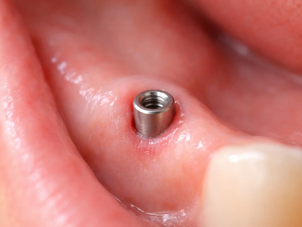
Does Gum Grow Around Implant? What to Expect and Fix
Explains if gum can grow around or over a dental implant, what’s normal healing, and how to address recession risks.
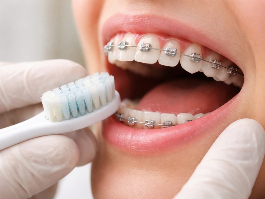
Why Do Gums Grow Over Braces? Causes and What to Do
Learn why gums cover braces and what to do now with hygiene steps, red flags, and orthodontist treatments.
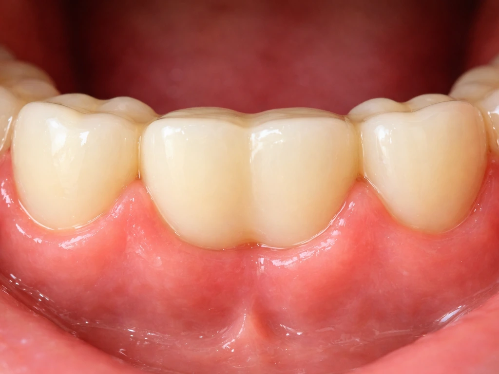
Do Gums Grow Around Bridge? What’s Normal and What’s Not
Learn if gums can grow around a dental bridge, what gaps mean, inflammation signs, and next steps to protect abutment te