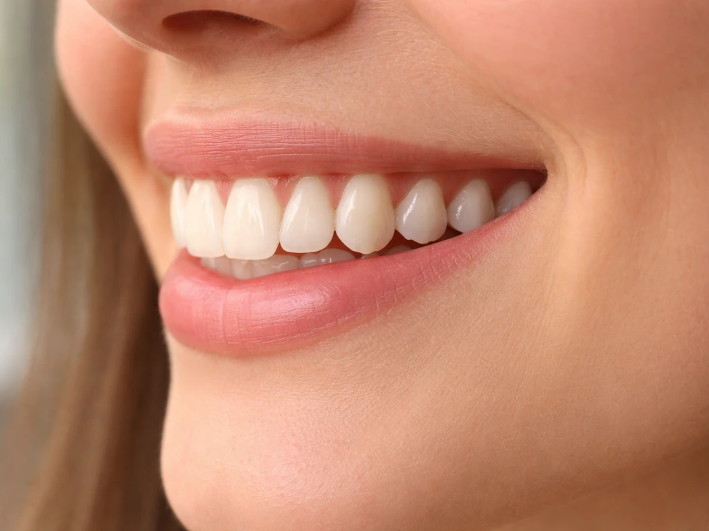
Adult teeth cannot grow longer the way a child's teeth grow in during development. In general, for adults there is no safe way to make teeth grow faster, and changes in “tooth length” usually come from gum recession or wear rather than new tooth growth make your teeth grow in faster. What you're actually seeing when a tooth looks longer than it used to is almost always gum recession, enamel wear, or a slow over-eruption process, not new tooth structure forming. The one real exception is kids and teenagers, whose permanent teeth are still erupting and can genuinely get taller as the jaw develops. For adults, if your teeth look longer, something is being lost, not gained, and that matters a lot for how you handle it.
Can Teeth Grow Longer? What’s Possible and What Isn’t

Marcus Holloway
19 May 2026
True vs. "Looks Longer" Causes of Longer Teeth
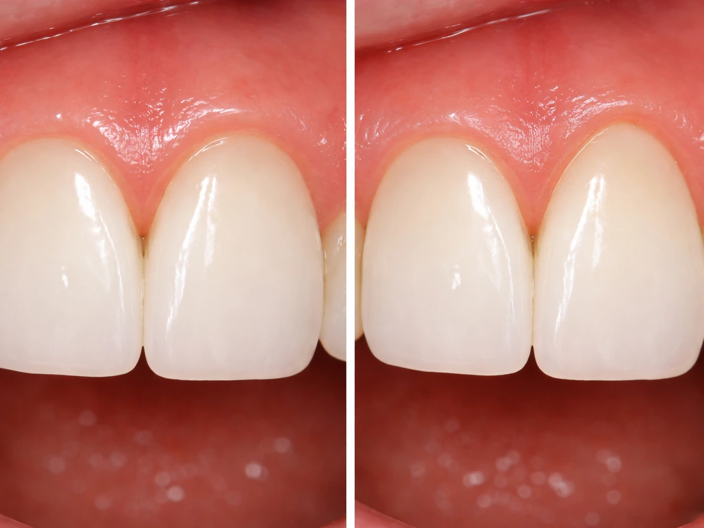
There's a real difference between a tooth that has genuinely changed its vertical position and a tooth that looks longer because gum tissue has pulled away from it. Both result in you seeing more tooth, but the cause and the fix are completely different.
True elongation can happen through a process dentists call continuous eruption or over-eruption. The Wikipedia overview of crown lengthening describes it as exposing more tooth by removing some gingival tissue and supporting bone, which is a clinical approach used when the longer-teeth appearance is due to needing more true tooth structure exposure blank" rel="noopener noreferrer">continuous eruption or over-eruption. Research published in the British Dental Journal confirms that teeth retain the ability to erupt throughout life, not just during childhood. A 10-year longitudinal study in adult women documented measurable changes in clinical crown height over time. A 16-year follow-up study also tracked how this continuous vertical movement interacts with periodontal changes. So yes, teeth do move vertically even in adults. But this is a slow, millimeter-scale shift, often triggered when there's no opposing tooth pushing back, like when a molar is extracted and the tooth above it slowly drifts downward. It's not the same as a tooth sprouting new structure.
The far more common reason teeth look longer is gum recession. The tooth itself hasn't moved much, but the gumline has dropped, exposing root surface that was previously hidden under tissue. You're not seeing more crown, you're seeing root. This matters because root surfaces are softer than enamel and more vulnerable to decay and sensitivity. The other "looks longer" culprit is tooth wear: when enamel or dentin erodes or grinds away at the biting edge, the shape of the tooth changes and it can look stretched or elongated even without any gum movement.
Age Matters: Childhood Eruption vs. Adult Changes
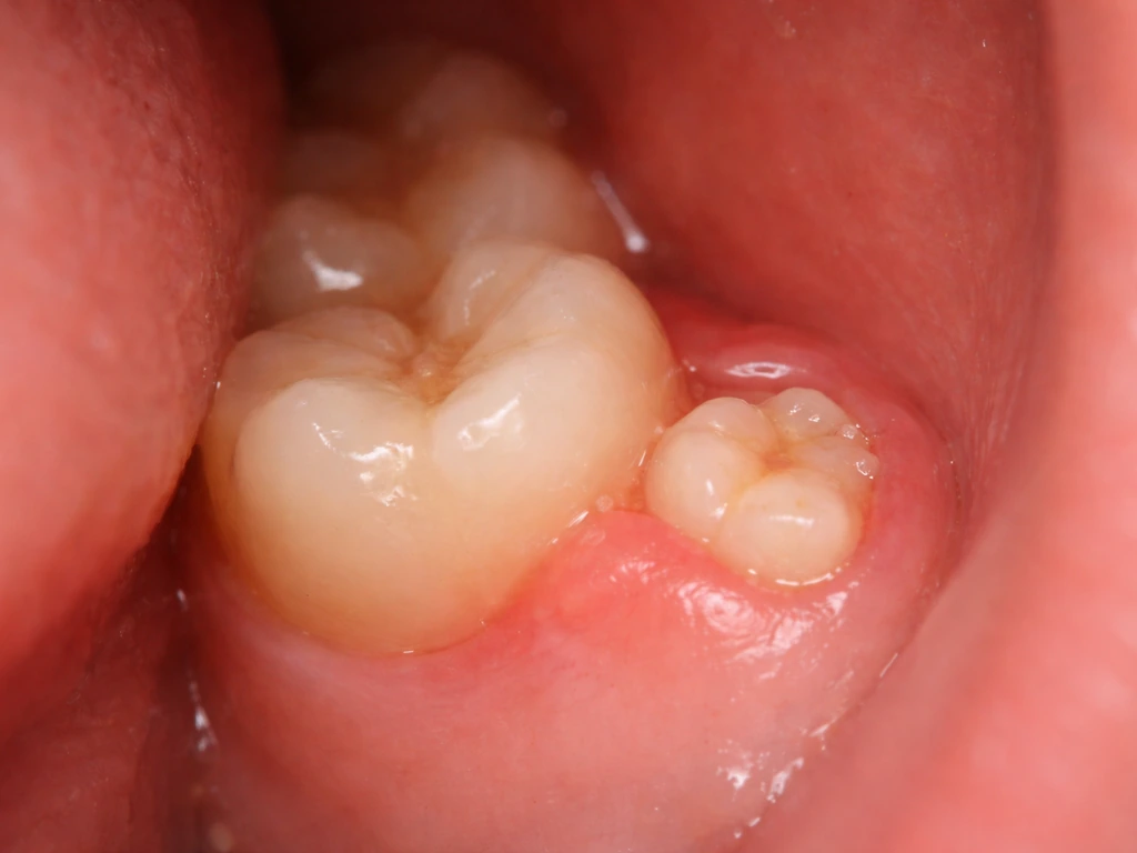
If you're a parent asking this about your child, the answer is much more straightforward: yes, their teeth are growing longer. Permanent teeth erupt gradually from around age 6 through the mid-teens. The incisors come in first, then premolars and canines, then second molars. Each of these teeth erupts in stages and continues to migrate into its final position over months to years. It's completely normal for a permanent tooth to look short or slightly sunken right after it breaks through and then gradually reach full height as the jaw grows. Wisdom teeth are a separate chapter, typically erupting between ages 17 and 25, and their arrival can shift how other teeth look and feel. If you’re wondering about wisdom teeth, keep in mind that their eruption can change how other teeth look, but correcting spacing or alignment usually takes an orthodontic evaluation. If you are wondering how to make wisdom teeth grow in faster, the most you can do is support healthy eruption and get checked early if pain or crowding suggests a problem.
For adults in their 20s, 30s, and beyond, the story shifts entirely. By the time you're out of your teens, eruption-driven tooth lengthening is essentially done. Any change you notice in tooth length from here on is almost always the result of the gumline moving down, the tooth wearing down, or a missing tooth allowing over-eruption of the opposing one. Age also increases your statistical risk: CDC and NIDCR data from large-scale NHANES surveys show that nearly half of adults over 30 have some form of periodontal disease, which is the number one driver of gum recession in adults. The older you are, the more likely the "longer tooth" you're noticing is a warning sign rather than normal development.
Gum Recession, Bone Loss, and Periodontal Disease Explained
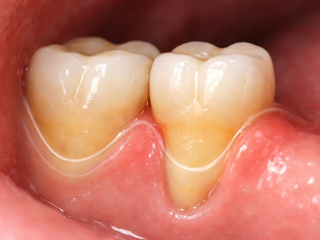
Gingival recession is the technical term for when the gum tissue that normally hugs the neck of the tooth migrates downward (or upward on lower teeth), exposing the root. Clinically, it's defined as an apical displacement of the gingival margin away from the cementoenamel junction, which is the natural boundary between crown and root. Once that root surface is exposed, you've lost a bit of protection that doesn't come back on its own.
Causes include periodontal disease, hard-bristled brushing technique, thin gingival tissue (some people are genetically predisposed to having very thin gum tissue, a "thin biotype," which multiple systematic reviews have identified as a significant risk factor), and orthodontic treatment that moves teeth outside of the bone envelope. The SDCEP guidance also highlights that tooth prominence in the arch and trauma from brushing are two of the most common everyday contributors.
Periodontal disease takes this further. Plaque-induced inflammation doesn't just move the gumline, it destroys the bone and the connective tissue (the periodontal ligament) that anchors the tooth. The ADA identifies the classic signs as deep pockets, bleeding on probing, gingival recession, and eventually tooth mobility. The AAP adds red or swollen gums and bleeding while eating. As the bone supporting the tooth is lost, the tooth can look dramatically longer, and it's genuinely at risk of being lost entirely if the disease isn't treated.
Enamel and Tooth Structure: What Can and Can't Regenerate
Here's where a lot of internet misinformation lives, so let's be direct. Enamel cannot grow back. Once the cells that build enamel (ameloblasts) are lost after a tooth fully erupts, they're gone. There is no biological mechanism for your body to regenerate enamel on a permanent tooth. Research published in RSC Biomaterials Science is clear: enamel repair after eruption is limited to surface-level remineralization, where minerals from saliva are incorporated into microscopic surface damage. That's not the same as regrowing bulk enamel. If you have significant enamel erosion, those surfaces are not coming back without a dentist.
Dentin is a different story. It has some limited repair capacity. When a tooth is injured or decay reaches a certain depth, odontoblasts (dentin-forming cells that stay active inside the tooth) can lay down a thin layer of reparative or reactionary dentin as a protective response. Research on pulp capping procedures confirms that a dentin bridge can form over an exposed pulp under the right conditions. This isn't regrowth in any meaningful cosmetic or structural sense, it's a defensive biological response. But it does mean the inner tooth has more repair capacity than the outer shell.
Gum tissue can partially recover from inflammation if the underlying cause is removed. Professional periodontal treatment can reduce pocket depth and gingival swelling, which sometimes makes the gumline look slightly better. True root coverage, however, requires surgery. Some periodontal regenerative procedures using guided tissue regeneration can even stimulate new cementum and periodontal ligament formation on previously exposed root surfaces, as documented in several clinical studies. But this is specialist-level treatment, not something that happens on its own.
The bottom line on regeneration: enamel gone is enamel gone. Some dentin repair happens naturally. Gum tissue can be surgically moved or grafted back into position. Lost bone can sometimes be partially regenerated with advanced techniques. None of this happens by waiting.
What Dentists Check: Symptoms, Measurements, and X-Rays
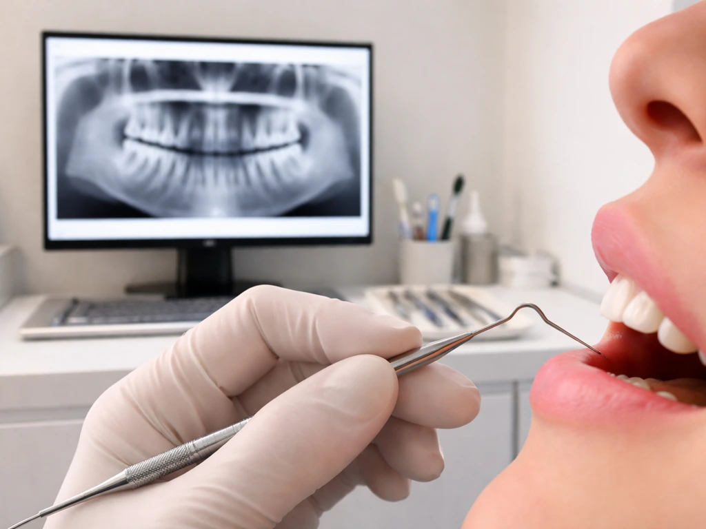
When you go to a dentist with the concern that your teeth look longer, they're running through a specific diagnostic checklist. Understanding it helps you know what to expect and what questions to ask.
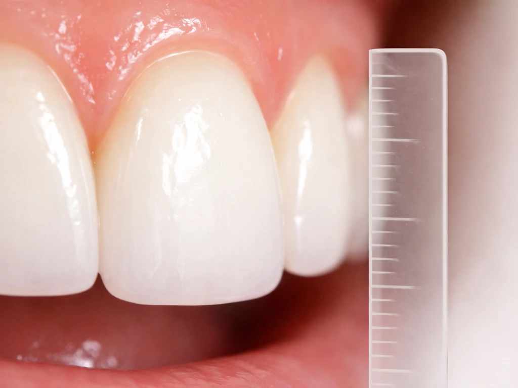
The first thing is a periodontal probing. A thin probe is walked around every tooth to measure pocket depth, the distance from the gum margin down to where the tissue attaches to the tooth. Normal is 1 to 3 millimeters. Pockets of 4 mm or more indicate a problem. Then they measure clinical attachment level (CAL), which accounts for both pocket depth and recession together, calculated from the cementoenamel junction rather than the moving gum margin. This gives a more accurate picture of true attachment loss. The ADA's measurement framework defines it this way: CAL equals probing depth plus the recession measurement from the CEJ to the gingival margin. This number tells you how much support the tooth has actually lost, regardless of where the gumline happens to be sitting.
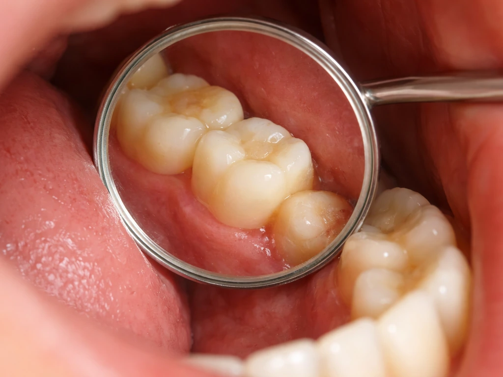
Next comes a visual recession assessment. Dentists often use Miller's classification (a widely used system based on where the gingival margin sits relative to the mucogingival junction and alveolar bone) to gauge severity and plan whether recession can be fully or only partially corrected. They'll also look at tooth wear patterns: incisal edge wear, notching at the gumline (abfraction), or flat, faceted surfaces that suggest grinding (bruxism). Research from Dentalcare notes that crown damage from wear can expose dentin and make teeth look longer even without any gumline movement.
X-rays are the final piece. Periapical X-rays show the full length of the root and the bone level around it. If bone has been lost to periodontal disease, the X-ray will show a lower bone crest than expected. Bitewing X-rays can show interproximal bone levels. For kids and teens, a panoramic X-ray can show which teeth are still developing and what stage of eruption they're in.
When to Worry: Urgent Signs to Act On Now
Most causes of "longer-looking teeth" are slow-moving and non-emergency, but a few symptoms mean you should get a dental appointment this week, not in a few months.
- A tooth suddenly looks visibly longer and feels loose or mobile
- You have bleeding gums every time you brush or eat, combined with bad breath that doesn't go away
- Sharp pain or throbbing at the root or jaw, especially with pressure
- A tooth has shifted position or there's a new gap between teeth
- You notice swelling at the gumline, especially around a specific tooth
- Significant new cold or hot sensitivity that appeared over weeks rather than years
- A child's permanent tooth is erupting but a baby tooth hasn't fallen out yet (retained primary tooth blocking eruption)
These signs can indicate active periodontal infection, an abscess, or rapid bone loss, all of which get worse with delays. The sooner they're addressed, the more options you have.
Treatment Options to Improve Tooth Length and Sensitivity
The right treatment depends entirely on what's driving the longer appearance. Here's how different causes map to different solutions.
| Cause | Primary Treatment | Can It Be Reversed? |
|---|---|---|
| Gum recession (early, non-periodontal) | Correct brushing technique, soft-bristle brush, desensitizing toothpaste | Partial improvement; full reversal needs surgery |
| Gum recession (significant) | Gingival graft (connective tissue graft or coronally advanced flap) | Often yes, especially Miller Class I/II |
| Periodontal disease with bone loss | Scaling and root planing (deep cleaning), possible periodontal surgery | Bone rarely fully regrows; disease halted |
| Enamel/dentin wear (bruxism) | Night guard, restorations (composite, crowns), occlusal adjustment | Worn enamel can't regrow; damage is restored |
| Acid erosion | Dietary changes, fluoride treatments, neutralizing acids, restorations if severe | Existing loss can't be reversed naturally |
| Over-eruption (missing opposing tooth) | Restoration/implant for missing tooth, occlusal adjustment, orthodontics | Partially correctable depending on degree |
| Crown exposure needed (cosmetic/restorative) | Surgical crown lengthening to expose more tooth structure | Deliberate procedure, not a reversal |
For sensitivity specifically, desensitizing toothpastes containing potassium nitrate or stannous fluoride are a reasonable first step if recession is mild and stable. They won't close the recession but they interrupt the nerve signaling that causes the sharp cold/sweet pain. In-office fluoride varnish or bonding agents applied over exposed root surfaces work faster and last longer. If the sensitivity is severe or the root surface is significantly exposed, a gum graft is both the aesthetic and protective solution.
On the surgical side, the coronally advanced flap and tunnel technique are both well-documented options for covering exposed roots. A systematic review comparing these two approaches found that the coronally advanced flap tends to offer higher mean root coverage for single recession defects. For more complex or widespread recession, connective tissue grafting from the palate is considered the gold standard. These procedures are typically done by a periodontist and have high success rates when the recession is caught before too much bone is lost.
If the problem is purely wear-driven, a custom night guard prevents further damage from bruxism. Composite bonding can rebuild chipped or worn incisal edges quickly and affordably. Veneers or crowns are considered when the wear is extensive and the tooth structure needs more comprehensive protection and restoration.
Self-Care and Prevention: Habits That Worsen vs. Protect
What you do at home every day has a bigger impact on whether your teeth continue to look longer than almost any clinical treatment. That said, if your goal is to make molars appear longer faster, you still need an evaluation to determine whether you are dealing with true eruption or gum recession teeth continue to look longer. If you want teeth to look longer, the most “natural” path is to stop the causes of loss like gum recession and enamel wear and then discuss appropriate dental options with a professional natural path. The research on this is consistent: traumatic toothbrushing with a hard-bristle brush is one of the top causes of recession, especially in people who already have thin gum tissue. A systematic review on orthodontic recession risk factors specifically named traumatic brushing and plaque accumulation as the leading causes of post-treatment recession. This doesn't mean brush less. It means brush correctly: a soft-bristle brush, gentle circular or modified Bass technique strokes, no scrubbing.
Here are the habits that make things worse:
- Brushing horizontally with pressure, especially at the gumline
- Using a medium or hard-bristle toothbrush
- Grinding or clenching teeth (bruxism), especially at night without a guard
- Drinking acidic beverages (sodas, citrus juices, sports drinks) frequently and swishing them around
- Acid reflux left untreated, which bathes teeth in stomach acid regularly
- Skipping flossing, which allows plaque to build up between teeth where the gum is most vulnerable
- Ignoring bleeding gums as "normal" when it's actually a sign of active inflammation
Here's what actually protects the gumline and tooth structure:
- Soft-bristle electric or manual toothbrush with gentle technique
- Fluoride toothpaste used twice daily to support surface remineralization
- Daily flossing or interdental cleaning to keep gum inflammation down
- A custom night guard if you know you grind your teeth
- Regular professional cleanings (every 3 to 6 months if you have a history of recession or gum disease)
- Staying hydrated and rinsing with water after acidic foods or drinks
- Treating acid reflux with your physician if it's a recurring issue
The BDJ review on tooth wear is emphatic that early identification of wear patterns and risk factors is essential because the damage is irreversible once it happens. That same principle applies to recession: catch it early, address the cause, and preserve what you have. Waiting to see if things get better on their own is rarely the right call with dental tissue, because unlike skin or muscle, the structures teeth are made of don't regenerate on their own. The good news is that with the right habits and the right treatment timing, you can protect what's there and, in many cases, restore what's been lost.
If you're curious about whether there are natural ways to support tooth eruption during development, the topics around how to make teeth grow in faster and how to make wisdom teeth grow in faster are worth exploring for age-specific guidance. Natural methods for supporting eruption focus on healthy development during childhood and teen years, not making adult teeth grow new structure faster how to make a tooth grow in faster. And if the concern is specifically about canine tooth length or molar eruption timing, those have their own nuances worth understanding separately. If your main goal is to understand how to make your canine teeth look longer, it usually comes down to whether you are dealing with normal eruption timing, gum recession, or tooth wear.
FAQ
If teeth can erupt throughout life, how do I tell if mine are truly over-erupting versus my gums receding?
Yes, but only within limits. In adults, “continuous eruption” can move teeth slowly, and the most noticeable cases are usually when a tooth has lost its opposing partner (for example, after an extraction) and can drift or over-erupt a bit. If you notice a rapid change, or multiple teeth look longer at once, it is more likely gum recession or bone loss than true eruption.
Can using a hard toothbrush make my teeth look longer, and what signs would suggest it’s recession from brushing?
Toothbrush hardness is not the only factor, technique matters too. Even with a soft brush, aggressive scrubbing along the gumline can worsen recession. A common pattern is wedge-shaped gumline wear plus sensitivity, especially near the canines or along the lower front teeth. A dentist or hygienist can map the pattern to determine the likely cause.
Will teeth whitening or enamel-whitening products make my teeth look longer or help the root show less?
Not usually. If the “longer” look is from gum recession, whitening products do not bring the gumline back or restore lost root coverage. Whitening can sometimes make the color contrast between exposed root dentin and enamel more noticeable, so the tooth may appear even more different until the recession is addressed.
If my teeth look longer due to wear, what can I do to stop it if enamel can’t grow back?
If enamel is worn, it cannot regrow, but you can reduce further loss. Typical options include fluoride varnish to strengthen remaining enamel, protective bonding for exposed areas, and bite-guard therapy if grinding is present. If you already have sensitivity, using a desensitizing toothpaste consistently and scheduling an exam helps prevent progression rather than “waiting it out.”
Can scaling and deep cleaning reverse gum recession, or is surgery usually required?
Yes. Periodontal therapy can reduce inflammation and swelling, which may improve how the gumline looks temporarily. However, true coverage of exposed root surface often requires procedures like guided tissue regeneration or grafting, depending on the recession type and the amount of lost attachment and bone.
If I have sensitivity when I drink cold water, does that confirm my teeth are growing longer from recession?
Sensitivity does not always correlate with the amount of visible “root.” Two people can have similar recession measurements but different discomfort depending on how much dentin is exposed, whether there are cracks from wear, and how inflamed the gums are. The practical next step is a periodontal assessment plus evaluation of bite and occlusion if wear is suspected.
Can missing teeth cause neighboring teeth to look longer, and does it change the urgency of replacement?
It can, especially if the “longer” tooth is drifting due to missing support in the bite or there is delayed replacement of the missing tooth. This is a reason dentists often recommend replacing teeth (with an implant or other option) rather than only monitoring. The effect is typically gradual, but it changes treatment timing.
What symptoms mean I should not wait and should see a dentist within days instead of months?
Common red flags include sudden worsening of recession, gum bleeding or swelling that is getting worse, pus, loose teeth, or pain with biting. These can indicate active periodontal infection or other acute problems, where earlier care can preserve bone support and limit how much root becomes exposed.
When I book an appointment for “my teeth look longer,” what should I ask the dentist to measure or evaluate?
Ask about the measurements they will use (probing depth and clinical attachment level), and whether the recession is stable or progressing. Also ask what category of recession they see, because prognosis differs by bone levels and tissue thickness. This helps you understand whether treatment is mainly desensitization, maintenance, grafting, or a combination.
Could orthodontic treatment or recent orthodontics make teeth appear longer, even if the gums aren’t receding?
Yes, especially if teeth are still erupting or shifting during adolescence. But in adults, many people confuse normal positional change with recession because teeth can also migrate slightly with orthodontics and bite changes. The most reliable way to separate them is an exam with standardized measurements and, when needed, updated X-rays.
If this started after I turned 18 or after a new dental issue, does that change whether it’s normal eruption versus gum problems?
Sometimes, and it is still worth checking. In young people, eruption can change crown height and how the smile looks over time. In adults, a sudden “taller tooth” look is more concerning for periodontal issues or bite-related over-eruption, especially if there is bleeding, inflammation, or rapid onset.
Next Articles
How to Make Teeth Grow Longer Naturally: What Works
Natural tooth lengthening vs gum recession explained, plus safe steps and when dentist care is needed
How to Make Wisdom Teeth Erupt Faster: Reality and Next Steps
Learn why wisdom teeth eruption timing varies, what affects it, and safe next steps for pain relief and dental care.
How to Make Wisdom Teeth Grow Straight: What Actually Works
Evidence based ways to manage wisdom teeth eruption, avoid complications, and what cannot truly “grow” straight