
You cannot make your canine teeth physically grow longer once they have fully erupted and your root development is complete. The biology is clear: adult enamel is acellular, meaning it has no cells left to produce more tooth structure, and the root is fixed in the jaw.
How to Make Your Canine Teeth Look Longer Safely

Marcus Holloway
28 May 2026
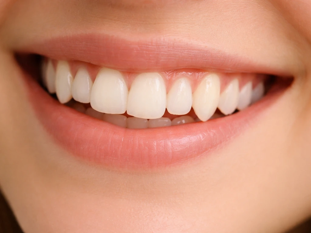
What you can do, depending on your age and situation, is encourage a canine that hasn't fully erupted yet to come in properly, correct one that's impacted or misaligned, reduce the gum tissue that's covering the tooth, or restore length that's been lost to wear. This connects to the question can teeth grow longer, because in adults the visible change is usually from restorative work rather than new tooth growth restore length that's been lost to wear.
All of those things can make your canine teeth look noticeably longer, they just work through different mechanisms than most people imagine.
Can canine teeth really grow longer, and what's actually possible at your age
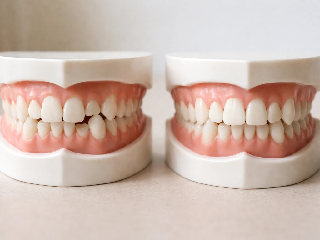
Canines go through two phases where visible length actually changes. The first is eruption: your permanent lower canines typically come in around ages 9 to 10, and [upper canines follow around ages 11 to 12. ](https://www. dentalcare.
com/en-us/ce-courses/ce500/permanent-dentition) Root development continues past that, usually finishing around age 13 to 15. During this window, the tooth is actively moving through the jaw and emerging through the gum, so if you're a kid or early teen with a canine that hasn't come in yet, there's a real conversation to be had about promoting healthy eruption.
The second phase where visible length can change is after eruption, and that one is mostly driven by how the gum sits around the tooth, not by the tooth itself getting longer.
Once you're an adult with fully formed canines, the tooth itself is done growing. No supplement, exercise, or technique changes that. What changes the appearance of tooth length in adults is gum position, wear, alignment, and whether any structural restoration is needed. Understanding which of those categories applies to you is the whole game.
Why your canines look short in the first place
Before you can fix something, it helps to know what's actually happening. There are a few distinct reasons a canine can look short or stubby, and they point toward completely different solutions.
The tooth hasn't fully erupted yet
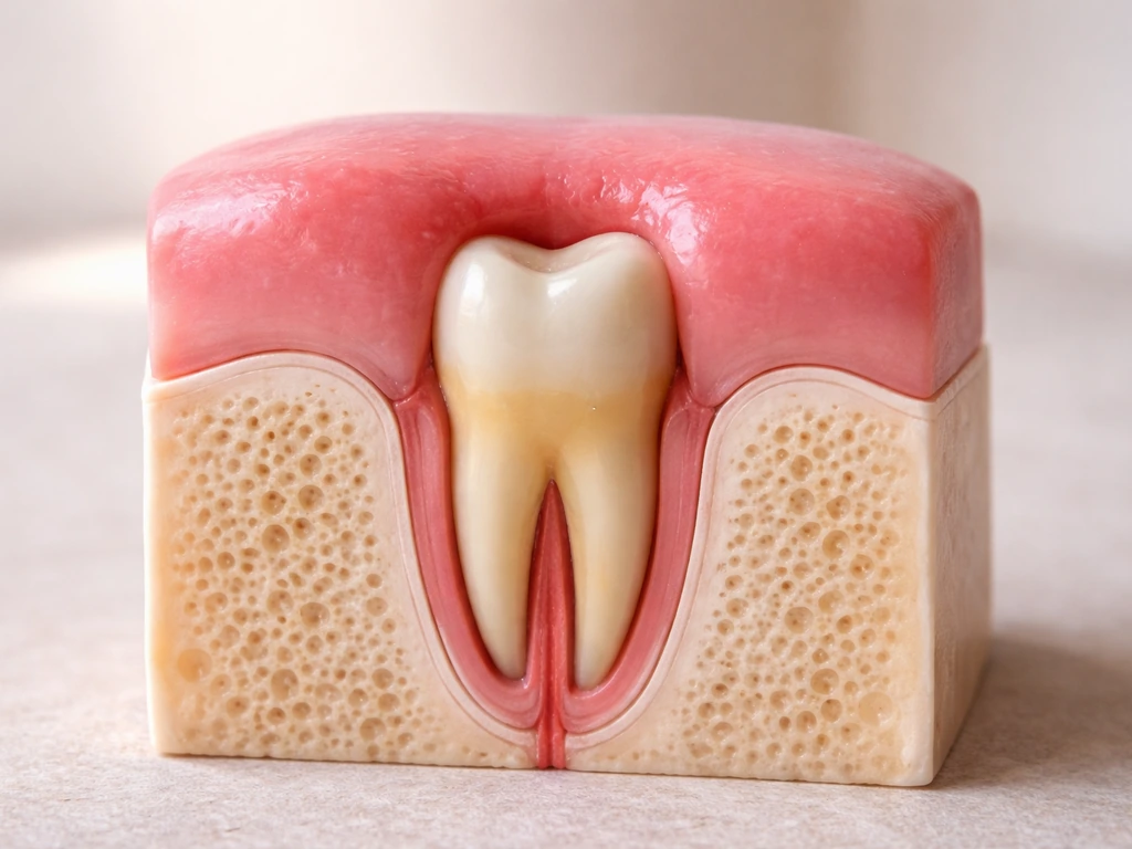
This is the most common cause in younger patients. Upper canines are actually one of the most commonly impacted teeth in the whole mouth, second only to wisdom teeth. Wisdom teeth can also get blocked and contribute to crowding, which is why impaction is a common theme when teeth do not erupt normally.
If the permanent canine is blocked by crowding, a retained baby tooth, or an unusual path through the bone, it either erupts partially or gets stuck entirely. The clinical tip-off in kids: if you can't feel a firm bulge on the gum just above where the upper canine should be by around age 10, that's worth flagging to a dentist.
Catching it early, ideally before the root is fully formed, gives orthodontic treatment the best chance of guiding it into place.
The tooth is fully erupted but covered by excess gum
Sometimes the canine has completely erupted, the crown is all there, but a thick band of gum tissue sits lower than it should, covering part of the tooth. This is called altered passive eruption and it's surprisingly common. The tooth isn't short, the gum is just in the way. A periodontist can address this with a minor gingivectomy procedure, and the change in visible tooth length can be dramatic.
Wear has shortened the tooth over time
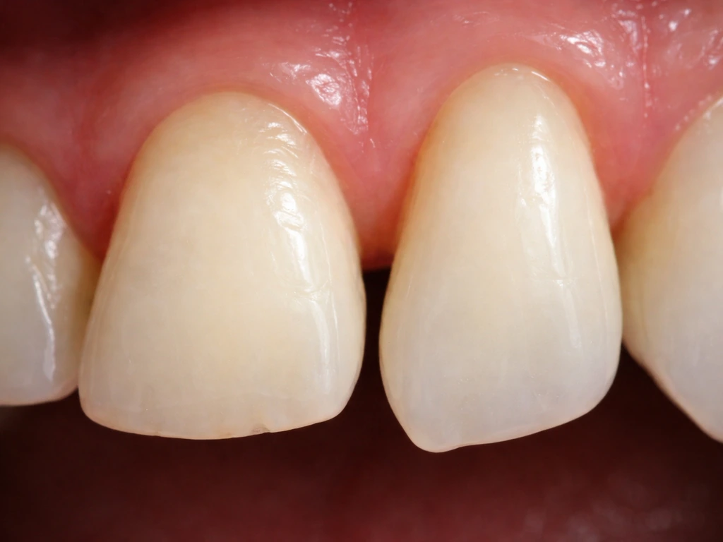
Bruxism (grinding), an uneven bite, or years of acidic food and drink can wear down the tip of a canine until it looks blunt and short. This isn't a gum issue or an eruption issue, it's structural loss, and restoring it means rebuilding what was ground away.
Gum recession has exposed the root but changed the appearance
Recession is the opposite problem: gum tissue has pulled back, exposing the root surface. This can make the tooth look longer in some cases, but it also creates sensitivity, root vulnerability, and an uneven color line where the crown meets the darker root. Left untreated, recession gets worse and threatens the whole tooth.
What 'natural' options can and cannot do
This is where most of the internet misinformation lives, so let's be direct. If you are wondering how to make your teeth grow, the key is understanding what can actually change (like eruption timing or gum position) versus what cannot (true enamel growth). You cannot stimulate your canine teeth to grow longer by eating certain foods, taking supplements, doing tongue exercises, or applying anything topically.
If your question is specifically about wisdom teeth, talk to a dentist or oral surgeon, because their eruption and growth follow different timing and biological limits than front teeth. Mature enamel has no cellular machinery left for self-repair or new growth, once the ameloblasts (the cells that built the enamel) complete their job during tooth formation, they're gone. Research is clear that biological bulk enamel regeneration is not achievable in humans yet.
Dentin, the layer under enamel, has slightly more regenerative potential in response to mild injury, but that process is internal, slow, and not something you can meaningfully influence from the outside.
What natural approaches can do is support the conditions for healthy eruption in kids. Good nutrition (particularly adequate calcium and vitamin D), removing obstacles like retained baby teeth on schedule, and maintaining low-inflammation gum tissue all help a developing canine follow a normal eruption path. These aren't hacks, they're just good dental health basics. For adults, the honest answer is that 'natural' lengthening isn't really a thing, but improving gum health, reducing grinding, and protecting existing tooth structure all prevent further shortening.
When to get checked, signs something isn't right
There are specific situations where you should stop waiting and get a professional evaluation. The earlier these are caught, the more options you have.
- A child is past age 10 and you can't feel or see any sign of the upper canine bulging through the gum
- A permanent lower canine hasn't appeared by age 11 or an upper canine by age 13
- The canine on one side of the mouth has erupted but the matching one on the other side hasn't
- Baby canines are still present well into the early teens
- The canine has come in but sits noticeably higher, lower, or more inward or outward than the teeth around it
- You notice a visible change in your bite, crowding near the canine area, or neighboring teeth shifting
- An adult canine looks progressively shorter over months or years (indicating ongoing wear or bite problems)
At a dental appointment for any of these concerns, expect a clinical exam plus radiographs. For straightforward cases, a panoramic X-ray gives a useful overview. If there's suspicion of impaction or the 2D images don't give enough detail about exactly where the tooth is sitting in the bone, cone beam CT (CBCT) is the gold standard, it provides three-dimensional positioning information that flat radiographs regularly miss, and it's what orthodontists and oral surgeons rely on when planning traction for impacted canines.
Treatment options that can actually increase visible length
Orthodontic eruption and traction for impacted or under-erupted canines
If a canine is impacted or only partially erupted, orthodontic traction is the evidence-based approach to bring it into proper position. The process typically involves braces or aligners to create space in the arch, followed by a minor surgical procedure to expose the impacted tooth and bond a small bracket or button to it. Elastic traction is then applied gradually to pull the canine into alignment. Total treatment time varies widely depending on how displaced the tooth is, the patient's age, and root morphology, studies report traction phases ranging from roughly 55 to 120 weeks, so this is a commitment. Younger patients with less complete root development generally respond faster.
Crown lengthening and gum recontouring
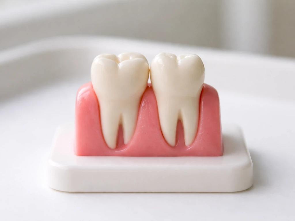
For adults whose canines are fully erupted but appear short because gum tissue covers too much of the crown, crown lengthening is a highly predictable procedure. A periodontist removes excess gum and sometimes a small amount of underlying bone to expose more of the natural tooth structure. The visible tooth gets longer without any change to the tooth itself. Results are stable long-term and this is one of the most straightforward ways to achieve a noticeably longer-looking canine in an adult.
Bite adjustments and occlusal changes
If an uneven bite is the reason a canine looks short, because it's being held out of full eruption by the opposing teeth, correcting the occlusion through orthodontics or selective adjustment can allow the canine to descend to its natural position. This is different from traction for impaction; it's more about removing the interference that's preventing full expression of available tooth length.
Periodontal and restorative options for protecting roots and rebuilding structure
Treating gum recession
If recession is exposing root surface below the enamel crown, the goal isn't to make the tooth longer, it's to get the gum back where it belongs to protect the root and restore normal aesthetics. The most effective approach for single-tooth recession is a coronally advanced flap combined with a connective tissue graft (CTG) taken from the palate.
Research shows this combination achieves complete root coverage in a substantial portion of Miller Class I and II recession cases, where the surrounding bone and tissue between teeth is still intact. Enamel matrix derivative (EMD) is sometimes used alongside surgical procedures to support periodontal regeneration of the structures that anchor the tooth. These procedures protect what you have, they don't add to tooth length, but they often restore the gum-to-crown ratio that makes the tooth look more proportional.
Restoring worn or shortened enamel
When grinding or acid wear has shortened the canine tip, the structural loss can be rebuilt with composite bonding or porcelain restorations (veneers or crowns, depending on how much tooth structure remains and what the bite situation is). Before any restoration, the underlying cause, usually bruxism or an acidic diet, needs to be addressed, or the new material will wear down just like the original did. A night guard for grinding and dietary changes work alongside restorative treatment, not instead of it. After rebuilding vertical dimension with restorations, the bite typically needs several months (roughly 6 to 9 months is a commonly cited adaptation period) to stabilize.
Forced extrusion for short clinical crowns
In some cases where the visible portion of the crown is genuinely short, perhaps due to fracture, severe wear, or subgingival decay, orthodontic forced extrusion is used to move the tooth and its surrounding bone and gum coronally, effectively creating more usable tooth structure above the gumline before a restoration is placed. This is a deliberate, controlled process coordinated between an orthodontist and a restorative dentist, and it's quite different from hoping a tooth will spontaneously grow. This is a deliberate, controlled process coordinated between an orthodontist and a restorative dentist, and it's quite different from hoping a tooth will spontaneously grow, which is closer to the question of how to make a tooth grow in faster.
Timeline, expectations, and how to move forward safely
Here's an honest breakdown of what different approaches actually look like in terms of timing and outcomes:
| Approach | Who it's for | Approximate timeline | Expected outcome |
|---|---|---|---|
| Orthodontic traction for impacted canine | Kids, teens, younger adults with unerupted canine | 55–120+ weeks of active traction, plus overall orthodontic time | Canine moved into arch; significant visible length gain |
| Crown lengthening / gum recontouring | Adults with excess gum coverage over fully erupted tooth | Single procedure; healing in 4–8 weeks | Immediate visible length increase; stable long-term |
| Gum recession surgery (CAF + CTG) | Adults with root exposure and recession | Single or staged procedure; 3–6 months full healing | Root coverage and restored crown-to-gum proportion |
| Composite bonding for wear | Adults with worn canine tips | 1–2 appointments | Restored length; requires bite assessment and maintenance |
| Porcelain veneer or crown for wear | Adults with moderate to severe structural loss | 2–4 appointments over several weeks | Durable length restoration; requires bite stability first |
| Forced extrusion for short clinical crown | Adults with subgingival fracture or severe wear affecting crown height | Several months of orthodontic movement before restoration | More tooth structure available for restoration; combined with other treatment |
A few things to actively avoid: skip any product or technique claiming to stimulate canine growth through topical application, diet alone, or 'natural elongation exercises', none of these are supported by dental biology, and some (like aggressive tongue pressure against teeth) can shift teeth in directions you don't want.
If you are tempted by claims on how to make your molars grow in faster, keep in mind that mature enamel cannot regenerate, and similar “growth stimulation” ideas are not supported by dental biology. Also avoid delaying evaluation if you or your child shows signs of impaction. The window for the simplest orthodontic intervention is widest before root development completes, which happens by the mid-teens for canines.
For example, a commonly cited dental-anatomy summary notes that permanent mandibular canines typically erupt around 9 to 10 years, with roots fully formed around 13 years. Waiting past that doesn't close all options, but it does make treatment longer and more complex.
Your practical next step depends on where you are. If you're a parent concerned about a child's canines, book a dental evaluation now, even a general dentist can take a panoramic X-ray and refer appropriately. If you're an adult who wants longer-looking canines, start with a consultation that includes an exam and photos so the provider can identify whether the issue is gum coverage, wear, or alignment.
Ask specifically whether a periodontist should be involved if there's any recession or gum tissue concern. If impaction is a factor at any age, ask whether CBCT imaging is warranted for proper three-dimensional localization before any treatment plan is finalized. Getting the diagnosis right first saves a lot of time and money on treatment that might not address the actual problem.
FAQ
How can I tell if my canine looks short because of gum coverage versus actual wear or structural loss?
You can measure “look” versus “real” tooth structure by having the dentist assess the crown display on photos (front-on and at an angle), then correlating that with gum probing depth and bite evaluation. If the tooth is shortened from wear or fracture, you will typically see abnormal crown contour and a history of grinding or diet exposure, whereas gum-based issues show normal crown anatomy but blocked visibility.
What age or signs mean I should stop waiting and get my child evaluated for an upper canine that looks short?
If a canine seems not to erupt as expected, the urgency is higher when you do not feel any firm gum bulge over the upper canine area by about age 10. Delays matter because orthodontic guidance is usually easier when root development is not complete, and treatment later often requires longer traction and more complex coordination.
Do I really need cone beam CT for an impacted canine, or is a regular X-ray enough?
Yes, CBCT can change the plan when 2D imaging is unclear, because it shows the exact position and angulation of an impacted canine relative to adjacent roots. This can affect whether the orthodontist can safely apply traction without risking damage to nearby teeth, and it helps decide whether additional surgical exposure steps are needed.
If I want a quick aesthetic fix, is it okay to do bonding or a veneer first?
Do not start with cosmetic bonding if the underlying issue is gum position or active grinding. A dentist should first address altered passive eruption, recession, or occlusal interference, because otherwise the new material may not match the gum line or it can be worn down quickly by the same forces that shortened the original tooth.
Will a night guard alone make my worn canine look longer again, or is restoration still needed?
Night guards help protect a rebuilt canine tip from further wear, but they usually should be fitted after a clinician confirms the bite and vertical dimension are stable enough. Also, a guard is not a substitute for addressing the cause of wear, for example frequent bruxism episodes, acidic intake patterns, or uneven contacts.
How do I know whether crown-lengthening is appropriate, or if I actually need gum recession treatment instead?
It depends on the diagnosis. Crown-lengthening can improve visible length when gum coverage is excessive, but it requires careful evaluation of periodontal support and “biologic width” to avoid long-term gum inflammation. If recession is the issue, the more appropriate goal is often root coverage rather than removing more tissue.
When would an orthodontist recommend forced extrusion for a short canine instead of bonding or a crown?
Orthodontic forced extrusion is different from “waiting for eruption” because it intentionally moves the tooth and its supporting tissues coronally under controlled conditions. This is typically coordinated so that restoration timing follows the tissue response, and it is chosen when you need more usable tooth structure above the gumline for a stable long-term restoration.
If my gum recession is making my canine look longer, what outcomes should I realistically expect from treatment?
For adult recession cases, the main decision is whether the root can be predictably covered. Your clinician should classify the recession (often using Miller Class I or II criteria), examine tissue and bone between teeth, and discuss expectations about sensitivity relief and color changes, since gum restoration improves appearance but does not “regrow” enamel.
Which at-home “canine lengthening” methods are most likely to be scams or could backfire?
Most “growth” products fail because mature enamel cannot be regenerated, and topical or exercise-based claims are not supported for producing additional tooth length. Be cautious with aggressive tongue or pressure techniques, since they can tip teeth, worsen alignment, or increase forces on the gums.
Can orthodontics make a fully erupted canine look longer even if there is no impaction or gum issue?
Yes, alignment can affect canine display even when the tooth itself is fully erupted. If the canine is held out of its natural position by a bite that interferes with eruption, correcting the occlusion can allow the tooth to express more of its crown without changing the tooth structure.
What red flags mean I should book sooner rather than waiting for a routine checkup?
If there is pain, swelling, or an abnormal eruption pattern, treat it as a reason for earlier evaluation. Also seek prompt care if you notice a tooth that appears stuck, a changing gum shape over time, or sensitivity that suggests recession, because intervention timing strongly influences options and complexity.
Next Articles
Can Teeth Grow Longer? What’s Possible and What Isn’t
See when teeth can look longer, what causes it, what can’t regrow, and next steps for sensitivity and gum recession.
How to Make Teeth Grow Longer Naturally: What Works
Natural tooth lengthening vs gum recession explained, plus safe steps and when dentist care is needed
How to Make Wisdom Teeth Erupt Faster: Reality and Next Steps
Learn why wisdom teeth eruption timing varies, what affects it, and safe next steps for pain relief and dental care.