Molars grow in the back of your mouth, behind the premolars, on both the upper and lower jaws. Your first permanent molars typically arrive around age 6 or 7, your second molars around ages 11 to 13, and your third molars (wisdom teeth) anywhere from 17 to 25. Once any of those molars are lost or extracted, they do not grow back. There is no third set of molars waiting in your jaw, and enamel does not regenerate on its own either. What you get is what you work with, so knowing exactly where they come in, when to expect them, and what to do if something goes wrong is genuinely useful information.
Where Do Molars Grow and When They Erupt

Marcus Holloway
6 Jun 2026
What molars are and where they sit in the mouth
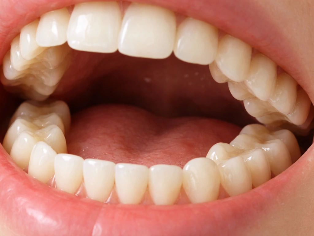
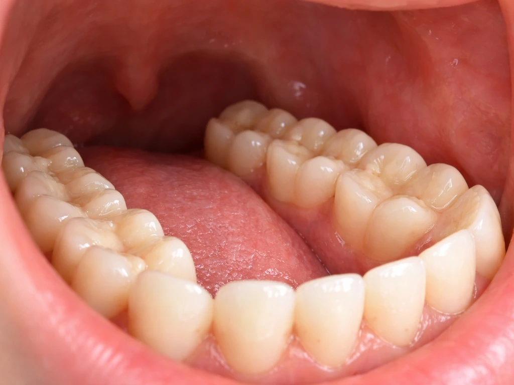
Molars are the large, flat-topped teeth at the very back of each side of your mouth. Their job is grinding and crushing food, and they are built for it, wider than any other tooth, with multiple cusps on the biting surface and multiple roots anchoring them into the jawbone. In a full adult mouth with all teeth present, you have 12 molars total: three on each side of the upper jaw and three on each side of the lower jaw. That includes your first molar, second molar, and third molar (the wisdom tooth) on each side.
Molars sit posterior to (behind) the premolars, which are themselves behind the canines and incisors. So if you run your tongue backward from the tip of your front teeth, you pass four incisors, then a canine, then two premolars, and then you hit the molars. The first molar sits right behind the second premolar. Each molar after that is positioned progressively further back, with the wisdom tooth being the rearmost tooth in the arch, when it erupts at all.
Exactly where molars erupt: upper vs lower, and the back-of-mouth pattern
Molars erupt symmetrically on both sides of both jaws. That means your first molar comes in on the upper left, upper right, lower left, and lower right, four teeth arriving around the same developmental window. The same pattern repeats for second and third molars. So when a parent asks "where does a molar grow in," the answer is always the same position bilaterally: behind the premolars, on both arches, both sides.
The upper and lower first molars often erupt at very similar ages, but there are subtle differences in timing across the set. Lower first molars tend to come in slightly before upper first molars. The upper second molars typically erupt a little later than lower second molars (around age 12 to 13 versus 11 to 12). Wisdom teeth can erupt upper and lower at similar ages, but the timing is highly variable and often asymmetrical, one side can erupt months or even years before the other, and some people only ever get one or two of the four.
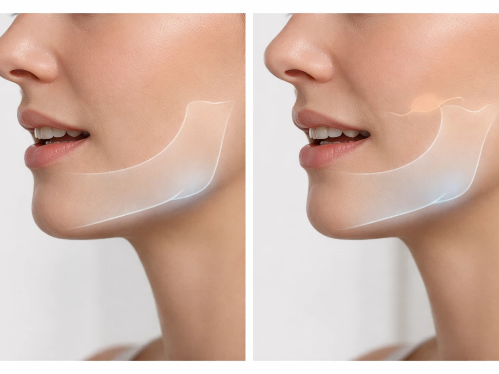
Molars do not push through a gap in the arch the way incisors do when baby teeth fall out. First and second molars erupt into newly created space as the jaw grows longer from back to front. The jaw physically extends to accommodate them, which is part of why questions about jaw growth and molar eruption are so closely linked.
Because the jaw grows longer as you age, that space is what allows some molars and related changes to happen in the teen years rather than later, such as the 20s jaw growth. Jaw growth is what creates the space that molars erupt into, which is why these teeth follow their own age windows as the jaw lengthens.
Third molars are the extreme version of this: they need space that often simply does not exist by the time they try to come in, which is the root cause of most impaction problems.
Eruption timeline: kids, teens, and wisdom teeth
Here is the standard eruption timeline for permanent molars. MedlinePlus notes typical permanent molar timing as first molars erupting around 6, 7 years, second molars around 11, 13 years, and third molars around 17, 21 years Here is the standard eruption timeline for permanent molars.. Keep in mind that variability is normal, kids can run a year or more ahead or behind these windows without it being a problem.
| Molar | Lower jaw eruption age | Upper jaw eruption age |
|---|---|---|
| First molar | 6–7 years | 6–7 years |
| Second molar | 11–13 years | 12–13 years |
| Third molar (wisdom tooth) | 17–21 years | 17–21 years |
The first permanent molars arriving around age 6 are sometimes called the "six-year molars." Many parents miss them entirely because there is no baby tooth to fall out first, these teeth erupt into brand-new space behind the primary teeth that are already there. Because they arrive so early and are so far back, they are also the molars most frequently affected by decay in childhood, which is one reason dentists often recommend sealants at that age.
Second molars, the "twelve-year molars," arrive during the middle school years. By this point the primary dentition is almost entirely replaced. Wisdom teeth are their own category. The Merck Manual and AAOMS both cite 17 to 25 as the realistic window, and that range is genuine, some people's wisdom teeth are fully erupted by 18, while others see movement at 24 or 25. blank" rel="noopener noreferrer">Third molars (wisdom teeth) are the last permanent teeth and typically appear between ages 17 and 21 years. A small percentage of people never develop one or more wisdom teeth at all, which is a normal genetic variation.
When "it hasn't come in yet" is normal vs a red flag
A molar that is a few months behind the typical eruption window is almost never a problem. What you are watching for are patterns that fall well outside the normal range or come with symptoms. For first and second molars, if the tooth has not appeared by a couple of years past the expected window and the baby tooth has already been lost, that deserves a dentist visit and an X-ray.
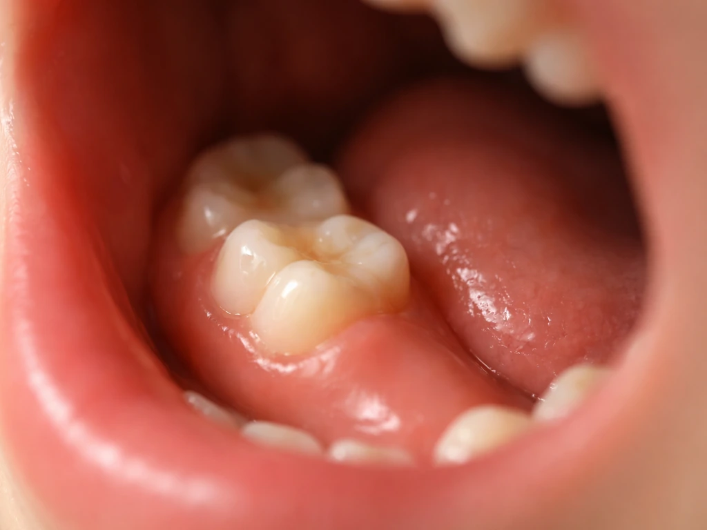
For wisdom teeth, delayed eruption or partial eruption is common and frequently the main story. A partially erupted wisdom tooth creates a tissue flap called an operculum over the biting surface, and that flap traps bacteria. The resulting infection, pericoronitis, causes localized pain, swelling, redness around the back of the mouth, and sometimes pus. It can become a persistent cycle if the tooth never fully erupts. This is not just normal soreness from a tooth coming in. If you have pain and swelling at the back of your mouth that comes and goes, that is a red flag worth acting on.
Other warning signs across all molar types include: a baby molar that stays put well past the expected age without any sign of a permanent tooth loosening it, visible displacement or tilting of a neighboring tooth (a sign of ectopic eruption), jaw pain that extends beyond mild soreness, or any sign of infection like swelling, warmth, or fever. These are the situations where the AAPD flags impaction, ectopic eruption, and ankylosis (where a tooth fuses to the bone instead of erupting) as clinical concerns requiring management.
Can a molar grow back after it's lost, damaged, or extracted?
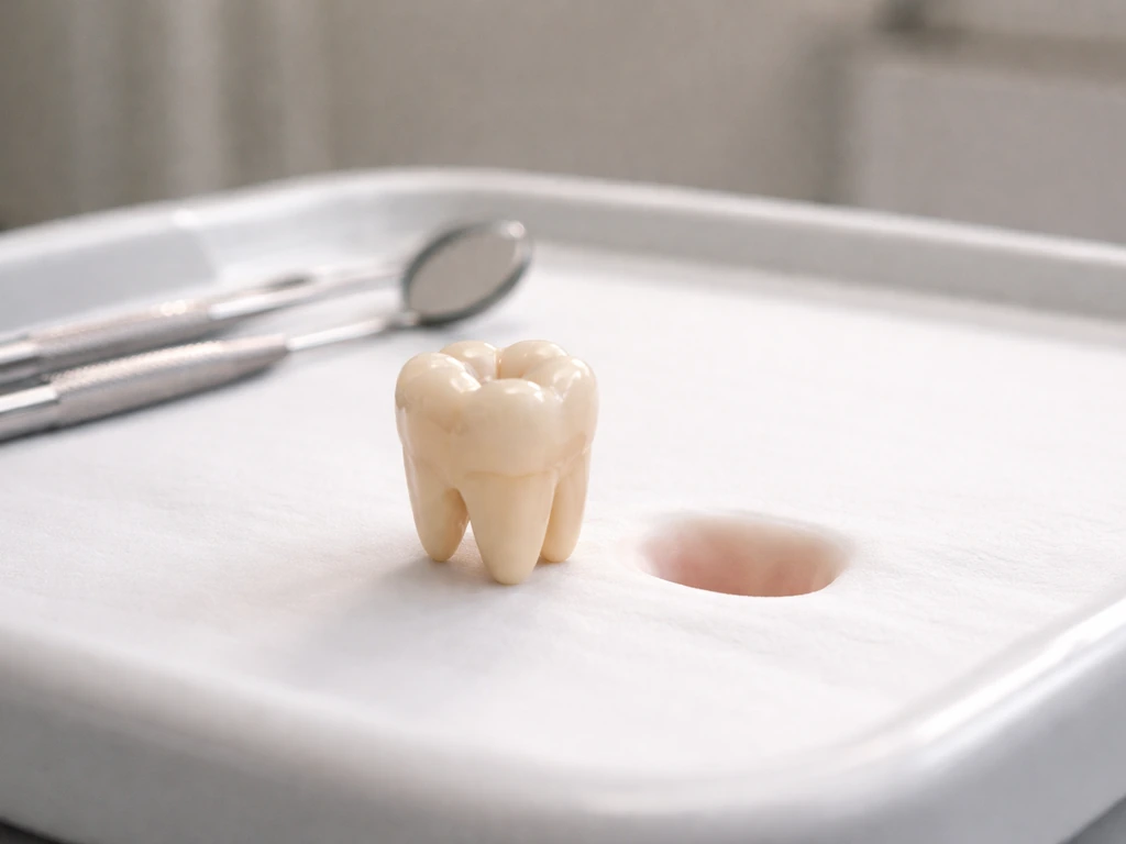
No. Once a permanent molar is fully erupted, there is no biological mechanism that will replace it if it is lost, extracted, or severely damaged. Humans are diphyodont, we get two sets of teeth, primary and permanent, and that is the end of the line. There is no third set waiting in the jaw. If a permanent molar is removed, that space is permanent unless it is filled with a dental prosthetic.
Enamel is in the same situation. Mature enamel is acellular, meaning it has no living cells after the tooth has fully formed. The cells that build enamel (ameloblasts) are shed when the tooth erupts. Once they are gone, the body has no way to rebuild enamel tissue from scratch. What is possible is remineralization: saliva contains calcium and phosphate ions that can repair very early surface mineral loss, which is why fluoride and good saliva flow genuinely help slow the very earliest stages of decay. But remineralization is patching microscopic mineral loss, not regrowing missing tooth structure. If a chunk of enamel is gone, or a cavity has created a hole, your body cannot fill it. That requires a dentist.
This is one of the most persistent myths in dental health: that cavities can "reverse" or that enamel can grow back if you eat the right foods or use the right toothpaste. Early-stage demineralization can be arrested and partially repaired, but a true cavity, a physical hole in the enamel and dentin, is not something your body can reverse on its own. The internet is full of this claim, and it is worth being direct: it is not how dental biology works.
How to self-check and what to ask your dentist
If you are worried about a molar that has not come in, the most useful thing you can do before a dentist visit is note your symptoms honestly. Is there any pain, and where exactly is it? Does it come and go or stay constant? Is there any visible swelling in the gum tissue at the back of your mouth? Is there a baby molar still present past the age when the permanent tooth should have appeared? Write this down, it helps the dentist narrow down quickly what is happening.
At the dentist, the key diagnostic tool is an X-ray. A panoramic radiograph (the kind where the machine rotates around your head) gives the dentist a full view of every tooth and root, including teeth still under the gumline. This is how impacted wisdom teeth are confirmed, how the position of an unerupted molar is assessed, and how a dentist decides whether a tooth will eventually erupt on its own or needs intervention. If a panoramic X-ray suggests that an impacted lower wisdom tooth is very close to the mandibular canal (the nerve canal running through the lower jaw), a cone beam CT scan may be recommended for a more precise 3D view before any surgery is considered.
Good questions to ask your dentist when you are concerned about a molar:
- Is there a tooth present in the X-ray that has not yet erupted, or is it simply missing?
- Is the unerupted tooth in a normal position, or is it angled, impacted, or blocked?
- Is there any sign of infection or damage to the surrounding teeth from the unerupted tooth?
- How likely is this tooth to erupt on its own, and over what timeframe?
- If it needs to be removed, what are my options for replacing that tooth afterward?
If a molar never erupted or was removed, here are your real options
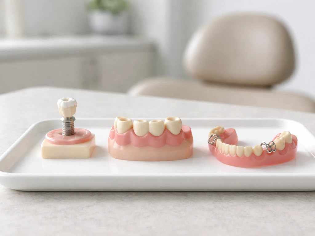
Since molars do not grow back, the practical question is what to do about the gap. The answer depends on which molar it is, where it is in your mouth, your age, and your overall dental health. For wisdom teeth specifically, many people do perfectly well without them and no replacement is necessary or recommended. For first and second molars, leaving the space empty is generally not a good long-term choice because neighboring teeth will drift and tilt over time, which can create bite problems and make future treatment harder.
The main restorative options after molar loss are dental implants, fixed bridges, and in some cases removable partial dentures. A dental implant is a titanium post placed into the jawbone that supports a crown, it is the closest thing to replacing a tooth root and is generally the longest-lasting option. A bridge uses the two neighboring teeth as anchors and spans the gap with a false tooth in between. Removable partial dentures are less common for single molar replacement but are sometimes appropriate depending on the overall situation.
There is also an interesting orthodontic option in some cases: if a wisdom tooth is impacted but otherwise healthy, an orthodontist can sometimes use it as a replacement by guiding it forward into the space left by a missing second molar. This is called molar protraction and it is not universally applicable, but it is a legitimate treatment path that avoids implant surgery for some patients. It is worth asking about if you are young and have a healthy unerupted third molar and a missing or damaged second molar.
If a child loses a primary (baby) molar early due to decay or injury, a space maintainer is often placed to hold that spot open until the permanent molar is ready to erupt. This prevents neighboring teeth from collapsing into the gap, which would block the permanent tooth from coming in correctly. This is one of the core reasons early childhood dental care matters so much, preserving space for the teeth that are on their way.
The bottom line is straightforward: molars grow in once, behind the premolars, on both arches, on both sides. They arrive in three waves across childhood and early adulthood. If they do not erupt normally, there are real diagnostic steps and treatment options available. But if they are lost, nothing in your biology will replace them, and that is reason enough to protect the ones you have.
FAQ
How can I tell a molar is coming in versus just gum irritation back there?
Tooth-eruption discomfort usually feels like mild pressure or soreness that improves over days, without spreading redness. If you notice swelling of the gum at the back of the mouth, bad taste, pus, fever, or pain that keeps recurring (especially around a partially erupted wisdom tooth), that points to an infection like pericoronitis and should be checked promptly.
If my child’s first molars are late, should we wait or book an X-ray right away?
A short delay of months is often normal, but if a first or second permanent molar is well beyond the typical window by about a couple of years and the baby tooth is already gone, ask for an exam and an X-ray. The key detail is whether the baby tooth is still present and whether symptoms or spacing issues are developing.
Do molars always erupt on both sides at the same time?
No. First and second molars tend to be fairly symmetric, but wisdom teeth are the most variable. It is common for one side to erupt months or years before the other, and some people develop only one or two wisdom teeth. However, asymmetry becomes more concerning if you have persistent swelling, drifting of neighboring teeth, or repeated pain.
Where exactly should I feel molars when I run my tongue or check my teeth?
The first molar is typically right behind the second premolar. As you move farther back, each next molar sits progressively posterior, with the wisdom tooth being the rearmost when present. If you cannot identify any molar bumps by the expected age range, that is a reason to confirm with an exam rather than guessing.
Can wisdom teeth cause problems even if they are not fully erupted?
Yes. Partially erupted wisdom teeth can trap bacteria under a gum flap (operculum), leading to pericoronitis. You can have symptoms that come and go because the flap can repeatedly irritate and harbor bacteria, so “it comes and goes” is still a warning sign.
What should I do if a permanent molar seems missing after the baby teeth are gone?
Treat it as a “needs confirmation” situation. Schedule a dental exam and ask whether a panoramic X-ray is appropriate, because the tooth may be impacted, tilted, or ectopic. Waiting without imaging increases the chance neighboring teeth shift and complicate future space and bite alignment.
If a molar is lost, is orthodontics ever enough without replacement?
Sometimes, but it depends on which molar is missing, the person’s age, and the health of the surrounding teeth and bite. For first and second molars, leaving the space empty often leads to drifting and tilting over time, so most plans involve replacement (implant, bridge, or in select cases a removable partial) or active space management.
Is a dental implant always possible after a molar is extracted?
Not always immediately. Implants generally require adequate jawbone development and good overall dental health, which is why clinicians often delay or stage treatment for younger patients. Your dentist will also consider neighboring teeth alignment and whether the gap can be managed conservatively while growth finishes.
Could a molar fail to erupt because it fused to the bone?
Yes. Ankylosis is one reason a tooth may not erupt normally even when it should, and it can affect how the surrounding bite develops. This is another scenario where X-rays are important, because visual exam alone cannot reliably distinguish ankylosis from simple delayed eruption.
What symptoms should send me to urgent dental care rather than waiting for a routine appointment?
Seek urgent care if you have significant swelling, difficulty swallowing or opening your mouth, fever, rapidly worsening pain, or signs of spreading infection. Also go sooner if you see warmth and redness at the back of the mouth along with feeling unwell, because infections around molars and wisdom teeth can escalate.
If I still have a baby molar, does that guarantee the permanent molar will come in?
Not necessarily. A baby molar persisting beyond the expected time can be a clue that the permanent tooth is delayed, impacted, or positioned incorrectly. The practical next step is to have the area examined and, if appropriate, obtain imaging to confirm where the permanent molar is and whether eruption is likely.
Next Articles
Molars Grow at What Age? Eruption Timeline for Parents
Molars growth timeline by type: when first, second and wisdom teeth erupt, what growth means, and when to see a dentist.
Does Jaw Grow in Your 20s? Growth, Teeth, and What to Do
Find out if the jaw truly grows in your 20s, how teeth and orthodontics change it, and what to do next.
When Molar Teeth Grow: Eruption Timelines and What to Expect
Molar eruption timelines, how tooth growth differs from regrowth, normal ages, delays, and when to see a dentist.